

Things Every Woman Should Ask Her Gynaecologist But Never Does

Things Every Woman Should Ask Her Gynaecologist But Never Does There’s a strange kind of silence that fills the room right before a gynaecology appointment ends. The doctor asks, “Any questions?” and most women say no, even when they have at least three things they’ve been meaning to ask for months. It’s not that the questions aren’t important. It’s that they feel awkward, embarrassing, or somehow too small to bring up. But gynaecologists hear these questions every single day. Nothing about your body is strange to them that’s literally the job. So here are the questions women quietly wonder about but rarely say out loud, and why asking them actually matters. 1. “Is My Period Actually Normal?” Most women have never compared notes on what a ‘normal’ cycle looks like, so they assume their experience heavy flow, irregular timing, severe cramps, is just how periods are. Sometimes it is. Often it isn’t. Cycle length, flow heaviness, clotting, and pain levels can all point to underlying conditions like PCOS, fibroids, or hormonal imbalances. There’s no harm in asking your doctor to simply confirm whether what you’re experiencing falls within a typical range. 2. “Why Does Sex Hurt Sometimes?” This question goes unasked more than almost any other, usually out of embarrassment. But pain during intercourse, whether occasional or consistent, has several possible explanations, ranging from dryness and infections to conditions like endometriosis. It’s a medical symptom, not a personal failing, and it deserves the same straightforward conversation as any other health concern. 3. “What’s Actually Normal Discharge vs. a Problem?” Discharge changes throughout the menstrual cycle, and most of that is completely normal. But changes in colour, smell, texture, or volume can be early indicators of infection. Many women either ignore these changes or feel too uncomfortable to mention them. A simple description during a check-up is often enough for a doctor to tell whether it’s nothing or something worth investigating further. 4. “Am I Too Young (or Too Old) to Worry About This?” Women in their twenties often assume certain conversations fertility, bone density, hormonal shifts are years away. Women approaching menopause sometimes assume it’s too late to ask about symptom management. Both assumptions get in the way of useful conversations. There’s no ‘right age’ to ask about your reproductive or hormonal health. Earlier conversations simply mean more options later. 5. “What Birth Control Option Actually Fits My Body?” Many women stick with whatever contraceptive method they started with years ago, even if it’s causing side effects, simply because switching feels complicated. The truth is, there are multiple options pills, IUDs, implants, injections, each with a different hormonal profile and suitability. A direct conversation about side effects, lifestyle, and future family planning can completely change which option makes sense. 6. “Should I Be Getting Screened for Anything Right Now?” Pap smears, HPV testing, breast examinations, these aren’t things that should only happen when something feels wrong. Routine screening catches problems early, often before symptoms even appear, which is precisely when treatment is most effective. Asking your doctor what screenings are appropriate for your age and history takes the guesswork out of preventive care. 7. “Is This Related to My Hormones?” Mood swings, fatigue, weight fluctuations, acne, hair thinning, women often deal with these in isolation, treating each as its own separate issue. Many of these symptoms trace back to hormonal patterns connected to the menstrual cycle, thyroid function, or conditions like PCOS. Mentioning multiple symptoms together, rather than addressing them piecemeal, often helps a gynaecologist spot the bigger picture faster. 8. “What Should I Actually Be Tracking?” Most women don’t track their cycles with any real consistency, and many don’t know what’s actually useful to track. Flow, pain levels, mood changes, and cycle length are all small data points that, together, give a doctor a much clearer picture than a single appointment ever could. Asking what to track and how turns a once-a-year conversation into something far more useful. Why These Questions Stay Unasked A few patterns show up again and again: Worry about sounding ‘dramatic’ over something that feels minor Limited appointment time, so questions get rushed or skipped Cultural hesitation around discussing reproductive health openly Simply not knowing that a symptom is worth mentioning at all None of these reasons make the questions less valid. Gynaecologists are trained specifically to discuss these topics without judgment, that’s the entire point of the consultation. Have a Question You’ve Been Holding Back? Sayee Specialty Hospital’s Obstetrics & Gynaecology department welcomes exactly these kinds of conversations – no question is too small or too awkward to bring up. Whether it’s about your cycle, contraception, screenings, or something that’s simply felt ‘off’ for a while, our specialists are here to listen first and explain clearly. You can book a consultation with our gynaecology specialists, or call us directly at 9 976 976 976 to schedule a visit. To learn more about our full range of women’s health services, explore our departments. Recent Posts edit post 7 Warning Signs You’re Losing Your Vision (That Most People Ignore) 7 Warning Signs You’re Losing Your Vision (That… edit post Sleep Deprivation : The Hidden Catalyst For Mental Disorders Sleep Deprivation : The Hidden Catalyst For Mental… edit post A Month By Month Guide To The Pregnancy Journey A Month By Month Guide To The Pregnancy… edit post Red Food Myth In Anaemia Treatment Red Food Myth In Anaemia Treatment There’s a…
7 Warning Signs You’re Losing Your Vision (That Most People Ignore)

7 Warning Signs You’re Losing Your Vision (That Most People Ignore) Most of us take our eyesight for granted. We go about our day reading, driving, scrolling through our phones, without stopping to think about how much we actually rely on our vision. But here’s the thing: most eye problems don’t show up overnight. They build quietly, over weeks or months, with signs so subtle that people dismiss them as tiredness, age, or stress. The problem? By the time vision loss becomes obvious, some of the damage may already be done. This post walks you through seven warning signs that your eyes are trying to tell you something signs that are commonly brushed aside, but shouldn’t be. 1. Blurry Vision That Comes and Goes Occasional blur is something most people chalk up to screen fatigue or a rough night’s sleep. And sometimes, that’s exactly what it is. But when blurriness keeps coming back even after rest, it’s worth paying attention. Fluctuating vision can point to early stage conditions like diabetes-related eye changes, dry eye syndrome, or shifting fluid pressure in the eye. None of these are emergencies on their own, but all of them benefit from early detection. If blurry spells are happening more than once or twice a week, get your eyes examined. Don’t wait for it to become constant. 2. Increased Sensitivity to Light Photophobia the clinical term for light sensitivity isn’t just uncomfortable. When it appears suddenly or gets progressively worse, it can signal uveitis (inflammation inside the eye), corneal damage, or even early glaucoma. People often wear sunglasses indoors or squint under normal lighting, assuming it’s just a quirk. Sometimes it is. But pairing light sensitivity with headaches, eye pain, or redness changes the picture considerably. 3. Seeing Halos Around Lights at Night If streetlights, headlights, or even the glow of a screen seem to have rings or halos around them, your eye’s lens may be struggling to focus light properly. This is one of the earlier signs of cataracts, a condition that progresses very slowly, often over years, before it begins significantly affecting sight. It’s also associated with poorly managed refractive errors and, in some cases, early glaucoma. Either way, it’s not something to normalise. 4. Floaters Especially When They Suddenly Multiply Seeing the occasional floater those tiny specs or threads that drift across your field of vision is normal. The vitreous gel inside the eye naturally changes over time, and mild floaters are part of that. What’s not normal: a sudden shower of new floaters, especially when combined with flashes of light. This combination can indicate a retinal tear or detachment, which is a genuine ocular emergency. Hours matter in those situations. 5. Narrowing Field of Vision (Tunnel Vision) Peripheral vision loss is one of glaucoma’s hallmark early signs and it’s particularly dangerous because it’s so hard to notice. Our brains are remarkably good at filling in visual gaps, which means people can lose a significant portion of their side vision without realising it. If you’ve started bumping into things at the edges of your path, misjudging distances when reversing a car, or feel like your visual field has quietly ‘shrunk’ these are red flags. Glaucoma is largely irreversible if caught late, but very manageable when identified early. 6. Difficulty Reading Small Text (Beyond Normal Ageing) Presbyopia the gradual loss of near-focus ability after the age of 40 is normal. Almost everyone experiences it to some degree. So reading glasses become part of life for many people, and that’s fine. What’s different is when standard reading glasses stop helping, when one eye seems significantly weaker than the other, or when text distortion or central blurring sets in. These can indicate macular degeneration, which affects the central part of the retina and can progress more quickly than people expect. Age-related macular degeneration (AMD) is one of the leading causes of vision loss in adults over 50. Early intervention genuinely changes outcomes here. 7. Eye Pain Paired With Nausea or Headache Eye pain on its own can have many explanations, a scratch on the cornea, eyestrain, sinus pressure. But when severe eye pain comes alongside nausea, vomiting, or a throbbing headache, it can be a sign of acute angle-closure glaucoma. This is a medical emergency. Intraocular pressure spikes rapidly and can cause permanent vision damage within hours if not treated. If you or someone you’re with experiences this combination, don’t wait and see how it feels in the morning. Why Do People Delay Getting Their Eyes Checked? There’s no single answer, but a few patterns come up repeatedly: Vision changes gradually, so there’s no obvious ‘moment’ that prompts action People assume decline is just part of getting older Eye care feels less urgent than other medical appointments Awareness of eye disease risk factors like diabetes or family history is still limited The reality is that many vision-threatening conditions are completely treatable when caught early. The difficulty is that ‘early’ often means before symptoms feel serious. Concerned About Your Eye Health? Sayee Specialty Hospital in Padappai offers specialist consultations for patients experiencing any of the symptoms described above. If you’ve been putting off an eye check or have noticed changes in your vision that don’t feel quite right, this is a good time to get a proper assessment. Our team of specialist doctors are available for consultations — book an appointment online, or reach us directly on 9 976 976 976. You can also explore the full range of specialist services available at Sayee — view all departments. Recent Posts edit post Sleep Deprivation : The Hidden Catalyst For Mental Disorders Sleep Deprivation : The Hidden Catalyst For Mental… edit post A Month By Month Guide To The Pregnancy Journey A Month By Month Guide To The Pregnancy… edit post Red Food Myth In Anaemia Treatment Red Food Myth In Anaemia Treatment There’s a… edit post Understanding Neck Pain Understanding Neck Pain Neck pain has become one…
Sleep Deprivation : The Hidden Catalyst For Mental Disorders
Sleep Deprivation : The Hidden Catalyst For Mental Disorders There’s a particular kind of pride that’s crept into modern culture around not sleeping enough. The five-hour night worn like a badge. The “I’ll sleep when I’m dead” mentality that gets quietly celebrated in workplaces and social circles as evidence of ambition, productivity, commitment. Getting by on minimal sleep has been reframed somewhere along the way as a personality trait rather than a health risk. It isn’t. And the consequences of treating it as one are accumulating in ways that mental health researchers have been documenting with increasing urgency. What Sleep Actually Does and What Happens When You Skip It Sleep isn’t passive downtime. It’s one of the most metabolically active periods in the entire 24-hour cycle, during which the brain performs maintenance work that simply cannot happen while it’s also managing waking life. During deep sleep, the glymphatic system the brain’s waste-clearance mechanism flushes out metabolic byproducts that accumulate during the day, including proteins associated with neurodegeneration. During REM sleep, the brain processes emotional memories, consolidates learning, and runs what amounts to overnight emotional regulation. Neural connections are strengthened and pruned. Stress hormones are regulated. The prefrontal cortex, responsible for rational thought, impulse control, and emotional regulation is restored to full function. Disrupt that process consistently, and the effects aren’t subtle. Sleep-deprived brains show measurably reduced activity in the prefrontal cortex and heightened reactivity in the amygdala, the brain’s threat-detection centre. The result is a nervous system that’s simultaneously less able to regulate emotional responses and more reactive to perceived threats. Minor frustrations feel catastrophic. Social interactions feel more fraught. The capacity to step back, reframe, and cope with daily stressors is genuinely diminished, not as a matter of willpower or attitude, but as a direct neurological consequence of insufficient sleep. The Bidirectional Relationship With Mental Health For a long time, the standard clinical assumption was that poor sleep was a symptom of mental health conditions, a consequence of anxiety, depression, and other disorders rather than a contributor to them. The evidence has shifted that understanding considerably. The relationship between sleep disruption and mental health is bidirectional. Yes, depression and anxiety disturb sleep. But disrupted sleep also independently increases the risk of developing depression and anxiety in people who were previously well. Insomnia isn’t just a side effect sitting passively alongside mental health struggles, it actively contributes to their onset and worsens their trajectory once they’ve developed. Studies following people over time consistently find that individuals with chronic insomnia have significantly elevated rates of developing major depressive disorder and anxiety disorders compared to those sleeping adequately. The causal pathway runs in both directions, creating feedback loops that can be genuinely difficult to interrupt once established. Poor sleep worsens mood. Worsened mood disrupts sleep further. The cycle compounds. Beyond depression and anxiety, chronic sleep deprivation is associated with increased risk of psychosis, even in people with no prior psychiatric history. Sleep loss amplifies paranoid ideation, increases unusual perceptual experiences, and in extreme cases of total sleep deprivation, can produce frank hallucinations. The mechanisms overlap with those seen in psychotic disorders, suggesting that sleep plays a role in maintaining the neurological coherence that keeps perception and thought organised. Emotional Regulation the Most Immediate Casualty The most immediately observable effect of insufficient sleep for most people, before any clinical threshold is crossed is emotional dysregulation. The capacity to manage emotional responses, maintain perspective, and resist reacting impulsively to provocations deteriorates measurably after even a single night of poor sleep. Positive emotions are blunted. The normal hedonic responses enjoyment of pleasurable experiences, enthusiasm for upcoming events, satisfaction with ordinary moments are dampened. Negative emotional responses are amplified. The same stressor that feels manageable after a good night’s sleep feels disproportionately overwhelming after a poor one. Irritability spikes. Empathy reduces. The ability to read social situations accurately diminishes. For people already managing existing mental health conditions, these effects compound on top of an already challenging baseline. For people without a mental health diagnosis, they represent a progressive erosion of psychological resilience that, sustained over months and years of chronic inadequate sleep, can contribute to the development of the very conditions they didn’t previously have. Why the Culture Around Sleep Makes This Worse The cultural glorification of sleep deprivation creates a specific problem: it removes the social permission to take sleep seriously. When insufficient sleep is treated as a productivity strategy rather than a health risk, people are less likely to protect their sleep, less likely to recognise the cognitive and emotional changes it’s producing in them, and less likely to seek help when the consequences become significant. Extended working hours, always-on digital connectivity, the blue light exposure from screens in the hours before sleep, the social norm of late-night entertainment, these environmental factors have collectively and substantially degraded average sleep quality and duration across the population. The average person today sleeps significantly less than their grandparents did, and it shows in mental health population data. Reclaiming good sleep in this environment requires treating it as a genuine priority rather than something that gets whatever hours are left after everything else is done. What Actually Helps For people dealing with temporary sleep difficulties disrupted sleep during a stressful period, difficulty winding down, inconsistent sleep timing, sleep hygiene improvements make a real difference when applied consistently. Consistent sleep and wake times, including on weekends, are the single most effective sleep hygiene intervention, they anchor the circadian rhythm and make falling and staying asleep easier. Reducing screen exposure in the 60 to 90 minutes before bed limits the blue light suppression of melatonin that delays sleep onset. Keeping the bedroom cool, dark, and reserved for sleep rather than work or screens strengthens the mental association between the sleep environment and sleepiness. Avoiding caffeine after early afternoon and limiting alcohol, which fragments sleep architecture even when it initially feels sedating supports deeper, more restorative sleep. For chronic insomnia
A Month By Month Guide To The Pregnancy Journey
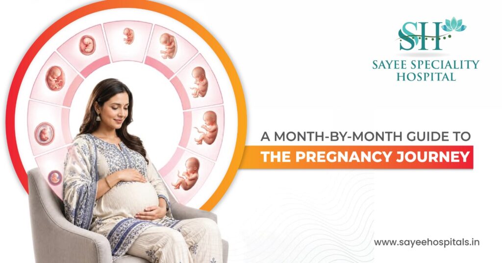
A Month By Month Guide To The Pregnancy Journey Forty weeks sounds like a long time until you’re living them. And then somehow it’s both endless and over in a blink, a period of constant change, occasional discomfort, moments of genuine wonder, and a running undercurrent of “is this normal?” that no amount of reading ever fully quiets. What helps, for most people, is understanding what’s actually happening not just to the baby, but to the body carrying it. Month by month, trimester by trimester, the pregnancy follows a sequence that’s been happening in human bodies for hundreds of thousands of years. Knowing what to expect at each stage doesn’t remove the uncertainty, but it does replace the vague anxiety of the unknown with something more grounded and manageable. TRIMESTER ONE: FOUNDATIONS OF LIFE (MONTHS 1- 3) Month 1 – Weeks 1 to 4 Most women don’t know they’re pregnant for much of the first month, which makes what’s happening during this period particularly remarkable in retrospect. Within days of fertilisation, the embryo implants in the uterine lining and the placenta begins forming. By day 22, the tiny heart still a simple tube at this stage begins beating. The neural tube, which will become the brain and spinal cord, starts closing. The embryo is roughly the size of a grain of rice, but the foundational architecture of an entire human being is already being laid down. For the mother, hormone levels shift dramatically and rapidly. Human chorionic gonadotropin – hCG rises steeply, triggering the hormonal cascade that sustains the pregnancy and produces the first symptoms. Breast tenderness and sensitivity are often among the earliest signs. Fatigue sets in quickly for many women not ordinary tiredness but a heaviness that makes it genuinely difficult to stay awake past early evening. Nausea may begin, typically worst in the morning though it can strike at any time. Month 2 – Weeks 5 to 8 The embryo transitions into a foetus during this period as the major organ systems begin forming heart, brain, liver, kidneys, lungs. The placenta becomes fully functional, taking over the task of sustaining the pregnancy. Eyelids form over the developing eyes. Fingers and toes begin to differentiate distinct digits with visible wrists and ankles. By the end of week 8, the foetus is around an inch long. Blood volume begins increasing, a process that continues throughout pregnancy and will ultimately result in the body producing around 50% more blood than usual. The expanding uterus presses against the bladder, and the increased kidney filtration rate means the need to urinate becomes noticeably more frequent. Nausea is often at its most intense during this month, and for some women the fatigue feels genuinely incapacitating. Month 3 – Weeks 9 to 12 The first trimester closes with the foetus reaching 2.5 to 3 inches in length and taking on increasingly recognisable features. Soft fingernails and toenails are growing. The first 20 tooth buds for the baby teeth that will appear years later are already present in the developing jaw. By the end of the first trimester, all the major organ systems are formed, even if not yet mature. The heartbeat is detectable with a handheld Doppler device, and most women have their first prenatal scan around this time. For many women, the end of the first trimester brings genuine relief nausea begins to ease, energy starts returning, and the risk of miscarriage drops substantially. Clothes begin feeling tight around the waist, though the bump isn’t usually visible to others yet. Blood pressure and hormonal fluctuations can cause headaches and occasional light-headedness, particularly when standing quickly. TRIMESTER TWO: GROWTH AND MOVEMENT (MONTHS 4 – 6) Month 4 – Weeks 13 to 17 The second trimester is, for most women, the most comfortable stretch of pregnancy. Nausea has typically resolved, energy has improved, and the bump isn’t yet large enough to cause significant physical discomfort. The foetus is now actively moving swallowing amniotic fluid, practising breathing movements, turning and repositioning though those movements aren’t yet strong enough to feel. The skin is transparent and pink, the umbilical cord fully functional, and the foetus reaches around 6 to 7 inches by the end of the month. The mother’s appetite usually returns properly during this period, and the visible baby bump begins to appear. The anatomy scan typically scheduled between weeks 18 and 22 – gives the first detailed structural picture of the baby’s development and is one of the most anticipated appointments of the pregnancy. Month 5 – Weeks 18 to 22 This is the month when most women feel foetal movement for the first time, a sensation described variously as fluttering, bubbles, or gentle tapping. As the pregnancy progresses those movements become more distinct and recognisable. The foetus is growing rapidly, establishing regular sleep and wake cycles, and developing the unique fingerprints and toe prints that will remain unchanged for a lifetime. The uterus has risen to roughly the height of the navel. The cardiovascular system is working significantly harder, the heart beats faster to manage the increased blood volume and some women notice palpitations or breathlessness during physical exertion. The anatomy scan during this window provides detailed assessment of fetal anatomy, placental position, and amniotic fluid levels. Month 6 – Weeks 23 to 27 By month six, foetal movement is well established and kicks are felt regularly. The eyes, previously fused shut, begin to open. The skin is red and wrinkled, covered with lanugo, the fine downy hair that helps regulate temperature before enough body fat has developed and vernix caseosa, a waxy protective coating. The baby weighs somewhere between 1.5 and 2 pounds and is around 12 inches long. The mother’s centre of gravity has shifted significantly by this point, contributing to the lower backache that becomes increasingly common in the second half of pregnancy. The round ligaments, which support the uterus, stretch as it grows, sometimes producing sharp, catching pains in the sides of the abdomen, particularly with sudden movements. Skin
Red Food Myth In Anaemia Treatment

Red Food Myth In Anaemia Treatment There’s a piece of nutrition advice that’s been passed down through families for generations, repeated by well-meaning relatives, and seemingly backed up by simple visual logic: eat red foods, because they look like blood, so they must build blood. Pomegranates, beetroot, red apples all commonly recommended to anyone diagnosed with anaemia or even just feeling a bit run down. It’s intuitive. It’s memorable. And it’s largely wrong. Why Colour Tells You Nothing About Iron Content The logic behind the red food myth rests on a kind of visual association that doesn’t hold up to actual nutritional analysis. The pigments that give fruits and vegetables their red colour anthocyanins in foods like pomegranate and red cabbage, betalains in beetroot are genuinely valuable compounds. They’re powerful antioxidants, they have documented anti-inflammatory properties, and they’re absolutely worth including in a healthy diet for reasons entirely separate from anaemia. But pigment and iron content are chemically unrelated. The compounds responsible for red colouration have nothing to do with how much elemental iron a food contains. A pomegranate gets its colour from anthocyanins. Its iron content roughly 0.3mg per 100g is genuinely modest, nowhere near enough to make a meaningful difference to someone with an active iron deficiency. Beetroot, despite its famously deep red colour and reputation as a “blood-building” food, contains around 0.8mg of iron per 100g again, a relatively small amount in the context of daily iron requirements, which sit around 8mg for adult men and up to 18mg for menstruating women. To put that in perspective: someone with iron-deficiency anaemia would need to eat an impractical quantity of beetroot or pomegranate daily to make any meaningful dent in their iron stores and that’s before accounting for how much of that iron the body actually absorbs. Where Red Foods Genuinely Help Just Not for the Reason People Think Here’s where the story gets more interesting, because some red foods do have a real role in anaemia management, but the mechanism has nothing to do with their colour or their own iron content. Strawberries, tomatoes, and red bell peppers are excellent sources of vitamin C. And vitamin C plays a genuinely significant role in iron metabolism specifically, it converts non-heme iron (the form found in plant foods) from its less absorbable ferric form into the more absorbable ferrous form, dramatically improving how much of that iron the body can actually take up. Some research suggests that vitamin C can increase non-heme iron absorption by several-fold when consumed alongside an iron-containing meal. So the connection is real it’s just indirect. These foods aren’t correcting anaemia by supplying iron themselves. They’re acting as absorption enhancers for the iron coming from other foods on the plate. A spinach salad with red pepper and a squeeze of lemon is doing something nutritionally meaningful. A bowl of pomegranate seeds eaten alone, on the assumption that it’s treating anaemia, mostly isn’t. Heme vs. Non-Heme Iron The Distinction That Actually Matters Understanding iron-deficiency anaemia properly requires understanding that not all dietary iron is created equal and this is the distinction that the red food myth completely sidesteps. Heme iron comes from animal sources red meat, poultry, fish, and organ meats like liver. The body absorbs heme iron remarkably efficiently, typically somewhere between 15% and 35% of what’s consumed, and that absorption rate isn’t significantly affected by what else is eaten at the same meal. Liver, in particular, is exceptionally iron-dense and remains one of the most efficient dietary sources of iron available. Non-heme iron comes from plant sources lentils, chickpeas, beans, spinach, fortified cereals, tofu. The body absorbs non-heme iron far less efficiently, somewhere between 2% and 20%, and that range is heavily dependent on what’s eaten alongside it. This is where the vitamin C pairing becomes genuinely useful, and it’s also where certain other dietary components work against absorption. Tannins and polyphenols found in tea, coffee, and red wine, bind to non-heme iron in the digestive tract and significantly reduce how much gets absorbed. This is well-documented enough that dietary advice for people managing iron deficiency commonly includes avoiding tea or coffee within roughly an hour of an iron-containing meal or supplement. Calcium, too, can interfere with iron absorption when consumed in large amounts at the same time as iron-rich foods. For people relying primarily on plant-based iron sources, whether by choice or circumstance these absorption factors aren’t minor details. They can be the difference between a diet that’s nutritionally adequate on paper and one that’s actually correcting a deficiency in practice. Why “Eating More Red Foods” Isn’t a Treatment Plan Anaemia isn’t a single condition with a single cause, and that’s really the core issue with any food-colour-based approach to managing it. Iron-deficiency anaemia is the most common type, but it’s far from the only one. Vitamin B12 deficiency and folate deficiency both cause anaemia through entirely different mechanisms affecting how red blood cells are formed rather than the iron available to make haemoglobin. Anaemia of chronic disease occurs when ongoing inflammation from conditions like rheumatoid arthritis, inflammatory bowel disease, or chronic kidney disease interferes with how the body uses iron, even when iron stores themselves are adequate. Anaemia from internal blood loss heavy menstrual bleeding, gastrointestinal bleeding from ulcers or polyps requires identifying and addressing the source of blood loss, not just replacing what’s being lost. Each of these requires a different management approach, and dietary iron intake red foods or otherwise addresses essentially none of them except straightforward dietary iron deficiency. Someone with B12 deficiency anaemia eating more pomegranates isn’t addressing the actual problem at all. What an Effective Approach Actually Looks Like Getting anaemia properly managed starts with diagnosis, not diet. A full blood count identifies whether anaemia is present and gives initial clues about the type, based on the size and characteristics of the red blood cells. Ferritin testing assesses iron stores specifically. B12 and folate levels rule those deficiencies in or out. Depending on the clinical picture,
Understanding Neck Pain

Understanding Neck Pain Neck pain has become one of those things that affects almost everyone at some point and increasingly, it’s affecting people younger than it used to. The combination of desk work, smartphones, and the kind of sustained sedentary posture that modern life demands has created a generation of people who know exactly what it feels like to reach the end of a workday with a neck that feels like concrete. Most of the time it resolves. But understanding why it’s happening, what’s actually going on in the cervical spine, and when it crosses the line from something manageable at home to something worth getting properly assessed, that knowledge makes a real difference. What’s Actually Causing It The cervical spine is a remarkable piece of engineering seven vertebrae supporting the full weight of the head, allowing rotation, flexion, and extension across a wide range, while simultaneously protecting the spinal cord and the nerve roots branching out to the arms. That combination of mobility and load-bearing makes it inherently vulnerable to both acute injury and cumulative strain. Poor posture is the most common driver in the modern context, and it’s worth understanding the mechanism rather than just accepting it as a vague explanation. When the head sits in a neutral position directly above the shoulders, the cervical spine bears roughly the weight of the head around 4 to 5 kilograms. As the head moves forward, which is what happens when you hunch over a laptop or look down at a phone the effective load on the cervical spine increases dramatically with each degree of forward tilt. At 45 degrees of forward head posture, the cervical spine is managing the equivalent of around 22 kilograms. Sustain that position for hours daily, and the muscles, ligaments, and discs at the base of the neck accumulate strain far faster than they can recover. Physical strain from repetitive movement, heavy lifting with poor mechanics, or sleeping in a position that leaves the neck unsupported for hours creates muscular tightness and, over time, joint irritation. Whiplash the rapid forced flexion-extension movement that occurs in rear-end collisions can damage muscles, ligaments, and facet joints in ways that sometimes produce symptoms for months after the initial injury. Age-related changes contribute increasingly from the fourth decade onward. Cervical spondylosis degenerative changes to the discs and vertebral joints, is almost universal in people over 60 on imaging, though it causes symptomatic pain in a smaller proportion. Disc herniation, where the soft inner material of a disc pushes through the outer layer and compresses an adjacent nerve root, produces the characteristic radiating pain, tingling, and weakness that can track down the arm in a dermatomal pattern. Spinal stenosis narrowing of the spinal canal itself, is a more advanced degenerative change that can compress the spinal cord as well as the nerve roots. Emotional stress deserves mention as a genuine physiological contributor rather than a psychological catch-all. The trapezius and other cervical muscles are among the first to tighten under sustained psychological stress, it’s an involuntary protective response, and people often carry significant muscular tension in the neck and shoulders for days or weeks during stressful periods without consciously registering it until the pain becomes noticeable. The Symptoms Worth Paying Attention To Neck pain itself varies considerably in character from a dull, persistent ache localised to the posterior neck, to a sharp, catching pain with specific movements, to a burning discomfort that builds through the day. Stiffness and reduced range of motion particularly difficulty rotating the head fully in one or both directions are common accompanying features. Where the picture becomes more clinically significant is when symptoms extend beyond the neck itself. Headaches originating at the base of the skull and spreading forward cervicogenic headache are a consistent feature of upper cervical dysfunction. Shoulder and upper back pain often accompany cervical muscle tightness, as the connected muscle groups react to the same underlying strain. Tingling, numbness, or shooting pain radiating down into one or both arms particularly if it follows a specific pathway toward certain fingers indicates nerve root involvement. This is cervical radiculopathy, and it warrants proper assessment rather than home management alone. Weakness in the hand or arm muscles alongside the sensory symptoms adds further urgency to getting that assessment done. How It Gets Diagnosed For most straightforward neck pain without neurological features, the diagnosis is clinical history, examination of range of motion, palpation of the cervical muscles and joints, and assessment of whether any neurological signs are present. Imaging is reserved for specific indications. X-rays identify bony changes degenerative changes, loss of disc height, facet joint arthritis, and in post-trauma cases, fractures. MRI provides the detailed soft tissue picture disc morphology, nerve root compression, cord involvement, and inflammatory changes that X-ray misses entirely. CT scanning offers superior bony detail and is particularly useful in acute trauma assessment. Blood tests are relevant when inflammatory or infectious causes are suspected raised inflammatory markers pointing toward conditions like rheumatoid arthritis or ankylosing spondylitis affecting the cervical spine. The important clinical flag is the combination of neck pain with upper limb neurological symptoms that combination consistently warrants imaging to characterise what’s compressing the nerve before a rehabilitation plan is designed. Management What Actually Helps For the majority of acute and subacute neck pain without neurological involvement, conservative management is effective and should be the starting point. Staying active matters more than most people expect. The instinct when the neck is painful is to rest and protect it, but prolonged immobility worsens muscular deconditioning and stiffness, and the evidence consistently supports gentle continued movement over rest. That doesn’t mean pushing through severe pain, but it does mean not treating the neck like it needs to be splinted. Physical therapy is the most evidence-based intervention for both acute and chronic neck pain. A physiotherapist assesses the specific pattern of dysfunction whether it’s primarily muscular, joint-related, or postural and designs a programme accordingly. Manual therapy targeting cervical joint mobility, combined with a progressive exercise
Understanding Bone Marrow Failure
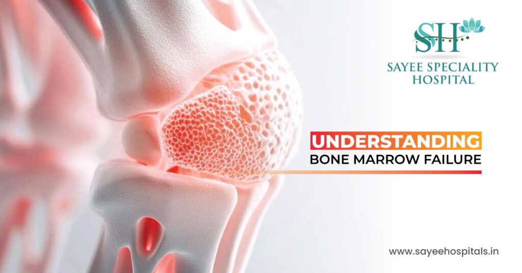
Understanding Bone Marrow Failure Most people don’t think much about their bone marrow until something goes wrong with it. It sits quietly inside the bones, doing one of the most essential jobs in the body manufacturing the blood cells that keep everything else functioning. When that process breaks down, the consequences reach into virtually every system, because almost nothing the body does works properly without a healthy, functioning blood supply. Bone marrow failure is serious. But understanding what it is, why it happens, and what modern treatment offers is a far more useful place to put your energy than fear alone. What Bone Marrow Actually Does Bone marrow is the soft, spongy tissue filling the inside of large bones the pelvis, sternum, vertebrae, and long bones. It’s where haematopoiesis happens the continuous process of producing new blood cells to replace the ones that die off daily in enormous numbers. Three cell lines come from this process, and each has a distinct job. Red blood cells carry oxygen from the lungs to every tissue in the body. Without enough of them, the body is starved of oxygen producing the fatigue, breathlessness, and pallor that characterise anaemia. White blood cells in their various forms are the immune system’s workforce, identifying and neutralising infections and foreign threats. Without adequate white cell production, the body loses its ability to defend itself even against organisms that a healthy immune system would handle without difficulty. Platelets are the clotting components small cell fragments that congregate at sites of injury and initiate the clotting cascade. Without sufficient platelets, bleeding becomes difficult to control and bruising occurs from minimal trauma. Bone marrow failure disrupts one, two, or all three of these cell lines simultaneously. When all three are affected, a condition called pancytopenia, the clinical picture is serious and the need for intervention urgent. Inherited vs. Acquired Two Very Different Starting Points Bone marrow failure can arrive through two entirely different pathways, and the distinction matters for how it’s investigated and treated. Inherited bone marrow failure syndromes are caused by genetic mutations present from birth passed down through families or arising as new mutations during development. Fanconi anaemia is the most well-known, a condition affecting DNA repair mechanisms that leads to progressive bone marrow failure alongside physical abnormalities and an elevated cancer risk. Diamond-Blackfan anaemia specifically affects red blood cell production, typically presenting in infancy with severe anaemia. Shwachman-Diamond syndrome combines bone marrow dysfunction with pancreatic insufficiency, affecting both blood production and digestion. These conditions require specialist haematological management from diagnosis and carry implications for family members who may be carriers. Acquired bone marrow failure develops during a person’s lifetime rather than being present from birth. The triggers are varied autoimmune attack on bone marrow stem cells is the mechanism behind aplastic anaemia, one of the most significant acquired bone marrow failure conditions. Viral infections including hepatitis viruses, HIV, and Epstein-Barr virus can damage marrow function. Blood cancers, leukaemia, lymphoma, multiple myeloma infiltrate the bone marrow and crowd out normal blood cell production. Chemotherapy and radiation therapy, while targeting cancer cells, inevitably cause collateral marrow suppression. Prolonged exposure to certain chemicals, pesticides, and drugs is associated with marrow damage in some cases. In a proportion of cases, no specific cause is ever identified idiopathic bone marrow failure that appears without a clear precipitating factor. Recognising the Symptoms The symptoms of bone marrow failure are largely the symptoms of what happens when the three blood cell lines become depleted and they can develop gradually or appear with surprising speed depending on how rapidly the marrow function is declining. Fatigue and weakness are almost universal. The kind of exhaustion that comes with significant anaemia isn’t the tiredness of a bad night’s sleep it’s a bone-deep, persistent depletion that doesn’t improve with rest. Breathlessness on minimal exertion, pallor, and a racing heart at rest are the more severe manifestations of significant red cell deficiency. Frequent infections bacterial, viral, and fungal that are unusually severe, prolonged, or recurrent signal white cell failure. Fever without an obvious source is a consistent feature. Infections that a healthy immune system would clear quickly become protracted and sometimes life-threatening when the white cell count is suppressed. Easy bruising, prolonged bleeding from minor cuts, nosebleeds, bleeding gums, and petechiae, the tiny, pinpoint red spots that appear under the skin when small blood vessels bleed without enough platelets to seal them indicate platelet depletion. Petechiae on the lower legs or around pressure points are particularly characteristic. Bone pain, particularly in the sternum, pelvis, or long bones, can occur when the marrow itself is under stress or infiltrated by abnormal cells. How the Diagnosis Is Made The diagnostic workup for suspected bone marrow failure starts with blood tests and builds toward direct assessment of the marrow itself. A full blood count is the starting point measuring the levels of red cells, white cells, and platelets simultaneously and identifying which cell lines are affected and to what degree. Reticulocyte count assesses how actively the marrow is attempting to produce new red cells. Blood film examination looks at the morphology of the cells present their size, shape, and appearance under the microscope provide diagnostic clues about the underlying cause. Vitamin B12, folate, and ferritin levels are checked to exclude nutritional deficiencies as a contributing factor. Viral serology tests look for the infections known to cause marrow suppression. Immunological testing investigates autoimmune causes. MRI of the spine and pelvis can assess marrow cellularity non-invasively, distinguishing between fatty, inactive marrow and normal haematopoietic marrow across different regions. The definitive investigation is bone marrow biopsy, a procedure where a needle is inserted into the posterior iliac crest under local anaesthesia to extract a core of bone marrow tissue for histological analysis. The biopsy establishes cellularity, identifies abnormal cell populations, and in combination with genetic and cytogenetic testing provides the specific diagnosis that drives treatment decisions. Treatment From Supportive Care to Transplant Treatment is tailored to the specific diagnosis, the severity of the failure,
Understanding Birthmarks in Babies
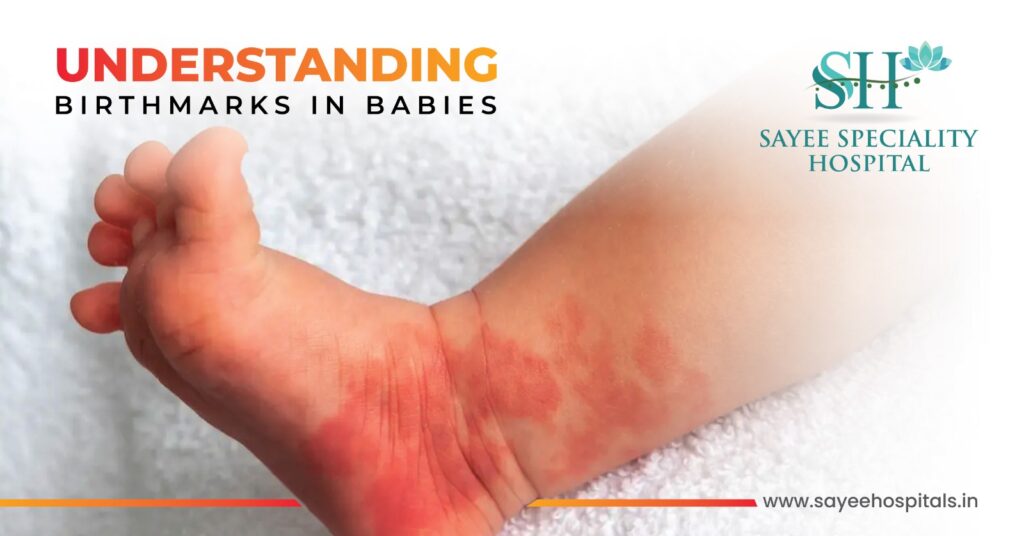
Understanding Birthmarks in Babies The first thing most parents do after a baby is born is look them over counting fingers, checking toes, taking in every detail of this new person. And sometimes, in that first examination, something unexpected appears. A reddish patch on the forehead. A bluish mark on the lower back. A raised strawberry-coloured lump that wasn’t there at the two-week check but has appeared by six weeks. Birthmarks are extraordinarily common more than one in ten babies is born with some type, and many more develop one in the first weeks of life. The vast majority are completely harmless. But knowing what you’re looking at, understanding which ones need watching, and recognising the small number of signs that warrant a doctor’s opinion that’s genuinely useful knowledge for any new parent to have. Two Categories Worth Understanding Birthmarks broadly fall into two types depending on what’s causing them, and that distinction matters because the two categories behave quite differently and have different implications for monitoring and treatment. Vascular birthmarks arise from abnormal blood vessels near the surface of the skin. Because blood vessels give them their colour, they tend to appear red, pink, or purple. Pigmented birthmarks arise from an excess of melanin, the pigment that determines skin colour concentrated in a particular area. These tend to appear brown, grey, or black. Within those two broad categories, the specific types have distinct characteristics, timelines, and considerations. Vascular Birthmarks Salmon patches are the most common birthmark of all, appearing in roughly half of all newborns. They go by the affectionate names “stork bites” when on the back of the neck and “angel kisses” when on the forehead or eyelids flat, pink or red patches that become more visible when the baby cries or gets warm. On the face, they almost always fade and disappear within the first couple of years. Stork bites on the nape of the neck can sometimes persist into adulthood, but they’re typically covered by hair and cause no problems whatsoever. No treatment needed, no monitoring required. Infantile haemangiomas commonly called strawberry marks are a different matter, though still reassuring in the majority of cases. They don’t usually appear at birth but develop in the first few weeks of life, growing rapidly through the first year before entering a slow, gradual phase of involution. By age five, around half have significantly faded. By age ten, most have resolved substantially. They appear as bright red, raised, soft lesions on the skin surface, though deeper haemangiomas develop beneath the skin and appear more bluish or skin-coloured with a slight swelling. Most haemangiomas require nothing more than observation and reassurance. The exceptions and they’re important are haemangiomas in specific locations or of significant size. A haemangioma near the eye can interfere with vision development if it’s large enough to obstruct the visual field. Haemangiomas around the mouth or nose can affect feeding or breathing. Large haemangiomas over the beard distribution of the face can occasionally be associated with internal haemangiomas affecting the airway. These warrant early specialist assessment and, where indicated, treatment typically with oral propranolol, a beta-blocker that has transformed the management of problematic infantile haemangiomas over the last fifteen years. Port-wine stains are flat, red or purple marks present from birth that don’t fade in fact they tend to darken and thicken over time if untreated. They’re caused by a localised malformation of capillaries in the skin and occur in roughly 0.3% of births. Unlike salmon patches and haemangiomas, they’re permanent without intervention. Port-wine stains affecting the face particularly around the eye and forehead — require medical evaluation because of an association with Sturge-Weber syndrome, a rare condition involving similar vascular malformations affecting the eye and brain. Pulsed dye laser treatment is the standard management approach and is most effective when started early. Pigmented Birthmarks Café-au-lait spots are smooth, flat patches of light brown pigmentation their name comes from the French for “coffee with milk,” which describes their colour well. They’re common, appearing in a significant proportion of children, and most are entirely benign. The clinical relevance comes with numbers, a single café-au-lait spot is almost always harmless. Six or more spots, particularly if they’re larger than a certain diameter, raise the possibility of neurofibromatosis type 1, a genetic condition that warrants investigation. A paediatric assessment for a child with multiple café-au-lait spots is appropriate. Dermal melanocytosis previously and less appropriately called Mongolian spots are flat, bluish-grey patches most commonly found on the lower back and buttocks, though they can appear elsewhere on the body. They occur when melanocytes, the pigment-producing cells, become trapped deeper in the skin during development. They’re particularly common in babies of Asian, African, Hispanic, and Middle Eastern heritage. They’re entirely benign and typically fade during early childhood without any intervention. The main clinical relevance is making sure they’re documented clearly at birth their bruise-like appearance has occasionally led to unnecessary safeguarding concerns when a healthcare professional unfamiliar with the mark encounters it later. Congenital melanocytic naevi are moles present at birth they can range from a few millimetres to, in rare cases, very large lesions covering significant areas of the body. Small and medium congenital naevi are common and generally low risk, though they warrant periodic monitoring for changes in appearance. Large and giant congenital naevi those covering a substantial surface area carry a small but elevated lifetime risk of melanoma and require specialist dermatological follow-up. Hair growing from a naevus is normal and doesn’t in itself indicate anything concerning. When to Get It Checked Most birthmarks need nothing more than a note in the baby’s records and periodic observation. But there are specific circumstances where a medical opinion shouldn’t wait. Any birthmark that grows rapidly — particularly in the first weeks of life deserves assessment to confirm it’s a haemangioma behaving normally rather than something requiring earlier intervention. Bleeding from a birthmark, persistent pain or tenderness, or ulceration of the surface are signs that the lesion needs review. A change
Thyroid Disorders And Irregular Periods

Thyroid Disorders And Irregular Periods Irregular periods get blamed on a lot of things stress, diet, over-exercising, coming off contraception. And sometimes those explanations are right. But there’s one underlying cause that gets missed surprisingly often, partly because it doesn’t announce itself obviously and partly because the connection isn’t widely known outside clinical settings. The thyroid gland a small butterfly shaped structure sitting at the base of the neck has a reach in the body that extends well beyond what most people associate with it. When it stops working properly, the ripple effects touch almost every system, including the one responsible for menstrual health. For a significant number of women with unexplained irregular periods, the answer isn’t gynaecological at all. It’s sitting in their thyroid function tests. What the Thyroid Does and Why It Matters for Periods The thyroid produces two primary hormones thyroxine (T4) and triiodothyronine (T3) that regulate the body’s metabolic rate. Essentially, these hormones set the pace for how every cell in the body uses energy. Heart rate, body temperature, digestive speed, cognitive function, mood all of these are downstream of thyroid hormone activity. But thyroid hormones don’t operate in isolation. They interact directly with the hormonal architecture of the reproductive system oestrogen, progesterone, and the pituitary hormones that regulate the menstrual cycle, FSH and LH. When thyroid hormone levels drift out of their normal range, that interaction becomes disruptive. The hypothalamic-pituitary-ovarian axis, the hormonal feedback loop that governs ovulation and menstruation is sensitive to thyroid status in ways that produce measurable, sometimes significant changes to the menstrual cycle. There are two directions thyroid dysfunction can go, and they produce opposite effects on periods. Hypothyroidism When the Thyroid Is Underactive Hypothyroidism occurs when the thyroid produces insufficient hormone. The most common cause is Hashimoto’s thyroiditis, an autoimmune condition where the immune system gradually damages thyroid tissue. It can also follow thyroid surgery, radioiodine treatment, or certain medications, and in some parts of the world iodine deficiency remains a significant cause. The systemic effects of hypothyroidism fatigue, weight gain, cold intolerance, constipation, dry skin, hair thinning, brain fog are well known. The menstrual effects are less widely understood but are a consistent feature of the condition. Women with hypothyroidism frequently experience heavy menstrual bleeding menorrhagia. Periods that soak through protection quickly, pass large clots, or last significantly longer than a week are common presentations. The mechanism involves the effect of low thyroid hormone on clotting factors and on the feedback signals that regulate the endometrial lining. Prolonged periods and increased frequency are also seen, the cycle shortening in some women so that periods come every two to three weeks rather than the usual interval. In more severe or longstanding hypothyroidism, periods can become infrequent or stop altogether, as the disruption to the hypothalamic pituitary axis becomes significant enough to suppress ovulation entirely. The broader symptom picture is useful for joining the dots. A woman presenting with heavy, prolonged periods who is also exhausted all the time, gaining weight without dietary changes, struggling with cold, and noticing her hair is thinning deserves thyroid function testing early in the diagnostic workup not as an afterthought once gynaecological causes have been excluded. Hyperthyroidism When the Thyroid Is Overactive Hyperthyroidism sits at the opposite end too much thyroid hormone, driving the body’s systems too fast. Graves’ disease, an autoimmune condition producing stimulating antibodies that activate the thyroid continuously, is the most common cause. Toxic nodular goitre and thyroiditis in its early inflammatory phase are other causes. The systemic picture is the reverse of hypothyroidism weight loss despite normal or increased appetite, heat intolerance, palpitations, anxiety, tremor, excessive sweating, and difficulty sleeping. The menstrual effects are also reversed. Hyperthyroidism tends to suppress menstrual flow rather than amplify it. Light periods, short cycles, infrequent periods, and amenorrhea complete absence of periods are the typical presentations. The high metabolic rate and the effects of excess thyroid hormone on oestrogen metabolism both contribute to a picture where the endometrial lining develops poorly and ovulation becomes irregular or absent. The cardiovascular symptoms of hyperthyroidism the racing heart, the palpitations, the anxiety often dominate the clinical picture and draw attention away from the menstrual changes. But for a woman of reproductive age presenting with light or absent periods alongside those systemic symptoms, hyperthyroidism should be high on the diagnostic list. The Fertility Dimension Thyroid dysfunction affects fertility through several overlapping mechanisms, all of which come back to the same central problem disrupted ovulation. Regular ovulation depends on the precise, coordinated hormonal signalling of the hypothalamic-pituitary-ovarian axis. Thyroid hormone imbalance interferes with that signalling at multiple points. In hypothyroidism, elevated TSH stimulates prolactin secretion, and elevated prolactin suppresses ovulation. In hyperthyroidism, altered sex hormone binding globulin levels change how oestrogen is metabolised and available, disrupting follicular development. The result is that both conditions reduce the frequency of regular ovulation and without consistent ovulation, conception becomes difficult. Studies examining women presenting to fertility clinics consistently find a higher prevalence of thyroid dysfunction than in the general population, and treating the thyroid condition frequently improves both ovulation regularity and conception rates. Beyond conception, untreated hypothyroidism during early pregnancy carries risks of miscarriage, preterm birth, and neurodevelopmental effects on the baby which is why thyroid screening is recommended for women with a history of thyroid dysfunction before and during pregnancy. Women with known Hashimoto’s thyroiditis who are planning to conceive should have their thyroid function optimised before trying. Other Conditions That Overlap One complication of the thyroid-menstruation connection is that several other common conditions produce similar menstrual symptoms, and they can coexist. Polycystic ovarian syndrome (PCOS) causes irregular, infrequent, or absent periods through a completely different mechanism insulin resistance and androgen excess rather than thyroid dysfunction. Endometriosis and uterine fibroids produce heavy, painful periods. Hyperprolactinaemia from causes other than hypothyroidism suppresses ovulation and periods. The practical implication is that a thyroid function test is an essential part of the workup for menstrual irregularity, but a normal thyroid result doesn’t end the
Comprehending Complex Regional Pain Syndrome (CRPS)
Comprehending Complex Regional Pain Syndrome (CRPS) Most pain makes a kind of logical sense. You injure something, it hurts, it heals, the pain fades. The injury and the pain exist in rough proportion to each other, and recovery follows a trajectory that feels predictable even when it’s slow. CRPS doesn’t work like that. It’s one of those conditions that genuinely challenges how most people understand pain, because the pain it produces is entirely disproportionate to whatever caused it, it doesn’t follow a healing curve, and it can persist and intensify long after the original injury has resolved. For the people living with it, that disconnect between what should hurt and what does hurt is one of the most isolating aspects of the condition. What’s Actually Going Wrong Under normal circumstances, the nervous system’s pain response is proportionate and self-limiting. Something damages tissue, the nervous system signals the brain, protective pain behaviour follows, the tissue heals, and the signal quietens. In CRPS, that shutoff mechanism fails. The nervous system either the peripheral nerves, the spinal cord, or the brain’s own processing centres, or some combination of all three becomes dysregulated in a way that sustains and amplifies pain signals far beyond what the original injury warrants. The current understanding describes CRPS as a disorder of neuroinflammation and central sensitisation. The nervous system essentially becomes hypersensitised calibrated to interpret ordinary sensory input as painful, and to produce pain responses that are wildly out of proportion to the actual stimulus. Touch that should feel neutral becomes agonising. A light breeze across the skin can be unbearable. The affected limb develops a life of its own in terms of temperature, colour, and autonomic behaviour changes driven by dysregulation of the sympathetic nervous system running alongside the pain pathways. CRPS is divided into two subtypes based on whether a specific nerve injury is identifiable. Type I previously called reflex sympathetic dystrophy occurs after an injury or illness that didn’t directly damage a named nerve. A wrist fracture, a sprain, a period of immobilisation, these are typical precipitating events. Type II, previously called causalgia, follows a confirmed nerve injury where the damage to a specific nerve is documented. The clinical presentation overlaps significantly between the two, and the distinction matters more for classification than for day-to-day management. It predominantly affects women, with onset most commonly in the fourth decade of life, though it can and does occur across a wide age range including in children and adolescents. What It Feels Like and the Symptoms Beyond the Pain The pain itself is described in terms that communicate how unlike ordinary pain it is. Burning is the most consistent descriptor deep, relentless, often described as feeling like the limb is on fire from the inside. Tearing, stinging, electric these appear frequently in patient descriptions. The intensity is severe enough that CRPS consistently scores among the highest on standardised pain scales, higher than many post-surgical pain states and comparable to active labour. Allodynia is one of the hallmark features pain triggered by stimuli that should cause no pain at all. Light touch, clothing against the skin, a change in air temperature, water from a shower. The threshold for pain drops so dramatically that the affected limb becomes almost impossible to tolerate normal contact with. Beyond the pain, the changes CRPS produces in the affected limb are striking. Skin colour shifts cycling between red, purple, and pale white, sometimes within the same day. Skin temperature changes, with the affected area often markedly warmer or cooler than the surrounding tissue. Skin texture can become thin, shiny, or mottled. Hair and nail growth on the affected limb may accelerate dramatically or slow to almost nothing. Swelling comes and goes. The limb can feel stiff, weak, and impossible to move normally partly because movement hurts, partly because the motor system becomes caught up in the same neurological dysfunction driving the sensory symptoms. All of these features the autonomic skin changes, the allodynia, the disproportionate pain are the nervous system’s dysregulation made visible. They’re not psychological. They’re not imagined. They’re the physiological consequences of a nervous system that’s lost its normal regulatory balance. What Triggers It In the majority of cases, CRPS follows a physical trauma, a fracture is the most common precipitant, with wrist fractures particularly over-represented. Surgery, sprains, crush injuries, burns, and even prolonged immobilisation of a limb have all been documented as triggers. The injury itself is often relatively minor and that’s part of what makes CRPS so disorienting. A fracture that would be expected to heal uneventfully in six to eight weeks instead becomes the starting point of a pain condition that can last years. Why some people develop CRPS following an injury while others with identical injuries don’t remains incompletely understood. Genetic predisposition, pre-existing differences in nervous system sensitivity, inflammatory response characteristics, and psychological factors have all been investigated as contributors. No single predictive factor has been identified. Getting a Diagnosis There’s no blood test, no imaging finding, no single investigation that confirms CRPS. Diagnosis is clinical reached through a combination of the history, the pattern of symptoms, and physical examination findings assessed against the Budapest Criteria, the internationally accepted diagnostic framework for CRPS. The Budapest Criteria require the presence of continuing pain disproportionate to the inciting event, alongside specific combinations of symptoms and signs across sensory, vasomotor, sudomotor, and motor categories. Meeting those criteria in the right clinical context, with other causes excluded, is how the diagnosis is established. Early diagnosis matters significantly. The longer CRPS goes unrecognised and unmanaged, the more entrenched the central sensitisation becomes and the harder it is to treat. A condition that might respond reasonably well to early aggressive rehabilitation becomes considerably more resistant after months or years of untreated neurological dysregulation. Treatment Rehabilitation First, Everything Else in Support Physical and occupational therapy are the foundation of CRPS management, and that priority reflects the underlying neuroscience. The goal isn’t just to manage symptoms, it’s to retrain the nervous system. Given that CRPS