

Ensuring A Healthy journey In High Risk Pregnancy

Ensuring A Healthy journey In High Risk Pregnancy The phrase “high risk pregnancy” lands differently depending on who’s hearing it. For some women it confirms something they’d already been quietly worrying about. For others it comes out of nowhere, a label attached to a pregnancy they’d assumed was straightforward. Either way, it tends to carry more fear than it needs to. High risk doesn’t mean high probability of a bad outcome. It means closer monitoring, more specialist involvement, and a more tailored care plan. Most women who carry a high risk pregnancy deliver healthy babies. Understanding what actually puts a pregnancy into that category and what that means in practice, is a far more useful place to put your energy than the label itself. What Actually Makes a Pregnancy High-Risk There’s no single definition with a neat checklist. High-risk is a clinical judgement that considers a combination of factors, some present before pregnancy, some that develop during it. Here’s what those factors generally look like. Age sits at both ends of the spectrum. Pregnancies in women under 20 carry elevated risks around premature delivery and low birth weight, partly linked to physical maturity and partly to socioeconomic factors associated with teenage pregnancy. At the other end, pregnancies in women over 35 often called advanced maternal age carry increased risks of chromosomal conditions like Down syndrome, gestational diabetes, placental complications, and hypertensive disorders. That doesn’t mean pregnancy after 35 is dangerous. Millions of women deliver safely and healthily in their late thirties and forties. It means the monitoring is more thorough. Pre-existing medical conditions form another major category. Chronic hypertension, type 1 or type 2 diabetes, obesity, thyroid disorders, epilepsy, heart conditions, autoimmune diseases, and poorly controlled asthma all have the potential to interact with pregnancy in ways that require careful management. Some conditions improve during pregnancy. Some worsen. Most require medication adjustments and more frequent specialist review. The key is that none of these automatically means a poor outcome, they mean the pregnancy needs more active management from the outset. Lifestyle factors matter more than many people expect. Smoking during pregnancy is associated with a significantly increased risk of miscarriage, placental abruption, preterm birth, and low birth weight. Alcohol carries risks of fetal alcohol spectrum disorder across the full range of consumption there’s no established safe level. Drug use, depending on the substance, creates its own spectrum of fetal and obstetric risks. These are modifiable factors, which makes them worth addressing directly and without judgment. Pregnancy specific complications can turn a previously low-risk pregnancy into a high risk one at any stage. Placenta praevia, where the placenta sits low and covers the cervical opening, requires careful monitoring and typically a planned caesarean. Fetal growth restriction, where the baby isn’t growing at the expected rate, signals that placental function needs close assessment. Rh sensitisation, where an Rh-negative mother develops antibodies against an Rh-positive baby’s blood cells, requires specific management to protect current and future pregnancies. Multiple pregnancies twins, triplets, or more, automatically carry higher risk. The uterus, placenta, and maternal physiology are managing more than one baby simultaneously, which increases the likelihood of preterm labour, growth discordance between babies, twin-to-twin transfusion syndrome in identical twins, and gestational hypertension. IVF pregnancies carry a higher rate of multiples than spontaneous conception, which partly accounts for their association with increased risk. Previous pregnancy history informs risk in the current pregnancy more than many people realise. A history of preeclampsia, preterm birth, stillbirth, recurrent miscarriage, or a baby born with a chromosomal or structural condition all raise the index of suspicion and influence how the current pregnancy is monitored and managed. How to Give a High-Risk Pregnancy the Best Chance The most important thing to understand here is that high-risk pregnancy management isn’t passive, it’s an active collaboration between you and your care team. The more informed and engaged you are, the better that collaboration works. Pre-conception planning is genuinely underutilised. If you have a known medical condition, a difficult previous pregnancy, or are in an age group that increases risk, a pre-pregnancy consultation with your GP or obstetrician before you conceive gives you the chance to optimise your health, review medications for pregnancy safety, discuss supplementation, folic acid at higher doses is recommended for some higher-risk women and understand your personal risk profile before it becomes urgent. That conversation is far easier to have before a positive test than after. Consistent prenatal care is the backbone of managing a high-risk pregnancy. The frequency and type of appointments will differ from a standard pregnancy more scans, more blood pressure monitoring, more frequent growth assessments. Women with complex medical conditions will often see a maternal-fetal medicine specialist alongside their regular obstetrician. Some will have input from cardiologists, endocrinologists, or haematologists depending on their specific condition. This level of involvement can feel overwhelming, but it exists because close monitoring catches problems early, when there are more options. Avoiding harmful substances is non-negotiable. If you’re still smoking, this is the time to access proper cessation support not just willpower. Alcohol should be stopped completely. Any medications, supplements, or herbal remedies should be reviewed with your doctor, because things that are harmless outside pregnancy aren’t always safe during it. Specialist Testing in High-Risk Pregnancies Beyond the standard antenatal scans and blood tests, high-risk pregnancies often involve additional investigations. Advanced ultrasound – including detailed anomaly scans and growth scans more frequently than the standard schedule – gives a clearer picture of fetal development and placental function over time. Biophysical profile assessments combine ultrasound findings with fetal heart rate monitoring to evaluate overall fetal wellbeing. For genetic risk assessment, cell-free DNA screening (sometimes called NIPT – non invasive prenatal testing) analyses fragments of fetal DNA in the mother’s blood to screen for chromosomal conditions like Down syndrome, with high sensitivity and without any procedural risk to the pregnancy. Where definitive genetic diagnosis is needed, amniocentesis – sampling the amniotic fluid – or chorionic villus sampling (CVS) – sampling placental tissue –
Cervicogenic Headaches: When The Headache Isn’t In Your Head

Cervicogenic Headaches: When The Headache Isn’t In Your Head Most people who get frequent headaches eventually start treating the head painkillers, dark rooms, cold compresses, caffeine. And for a lot of headache types, that’s a reasonable approach. But there’s a subset of headache sufferers who do all of that, consistently, and never quite get on top of it. The headaches keep coming back, the pain relief is temporary at best, and nobody seems to have a satisfying explanation for why. For some of those people, the problem was never in the head to begin with. The Neck as the Real Culprit Cervicogenic headache is classified as a secondary headache meaning it’s a symptom of something else rather than a condition in its own right. That something else is a structural problem in the cervical spine, specifically the upper neck vertebrae most commonly the C1, C2, and C3 levels. The mechanism is referred pain. The upper cervical spine and the head share overlapping nerve pathways, particularly through the trigeminal nucleus, a structure in the brainstem where signals from the face, scalp, and upper neck all converge. When there’s irritation or dysfunction at those upper cervical joints, the brain interprets that signal as coming from the head. The pain feels like a headache. It presents like a headache. But the source is in the neck, not the skull. What causes that cervical dysfunction in the first place varies. Whiplash from a car accident is one of the more common triggers. Cervical spondylosis, the degenerative joint changes that come with age and wear, is another, which partly explains why cervicogenic headaches tend to show up more frequently after the age of 30. Disc herniation, facet joint arthritis, and pinched cervical nerve roots are all documented sources. Sometimes it follows a sports injury. Sometimes it develops gradually without any clear incident. What It Actually Feels Like This is where cervicogenic headache earns its reputation for being difficult to diagnose, because on the surface, it can look almost identical to a migraine or a tension-type headache. The pain is typically one-sided, which is a useful clue. It usually starts at the base of the skull. the suboccipital region and spreads forward, often tracking toward the eye or temple on the same side. Some patients describe it as a pressure behind the eye that seems to have no obvious cause. The one-sided, eye-involving quality is what frequently triggers a migraine workup, and it’s not uncommon for people to carry a migraine diagnosis for years before anyone looks at the neck. The distinguishing features, when you know what to look for, are mechanical. The headache worsens with specific neck movements or sustained postures, looking down at a phone for a long period, turning the head a certain way, or holding the neck in one position during sleep. Limited cervical range of motion is a consistent finding on examination. Pressing on specific points in the upper neck the suboccipital muscles, the upper facet joints, often reproduces or intensifies the headache, which is a meaningful clinical sign. One detail that catches people off guard: the neck itself doesn’t always hurt. Patients frequently present with a headache and no significant neck pain, which removes the most obvious pointer toward a cervical source. That disconnect contributes to how long the diagnosis takes to arrive. Getting the Diagnosis Right There’s no single test that confirms cervicogenic headache, diagnosis involves piecing together the clinical picture from multiple sources. The International Classification of Headache Disorders criteria (ICHD-3) provide a diagnostic framework, requiring evidence of a cervical lesion or disorder known to cause headache, alongside clinical signs implicating the neck as the source. In practice, this means a detailed history, a hands-on physical examination assessing cervical mobility and tenderness, and imaging. X-rays, CT scans, and MRI are all used primarily to identify structural contributors like disc herniation, osteophytes, or facet joint degeneration, and to rule out more serious pathology like fractures or tumours. But here’s the important caveat: a normal scan doesn’t rule out cervicogenic headache. Imaging captures structure, not function. A cervical joint can be generating pain through abnormal movement or irritation without showing anything remarkable on an MRI. That’s why the physical examination and clinical assessment carry so much weight alongside the imaging findings. Diagnostic nerve blocks — injecting local anaesthetic around the suspected cervical source are sometimes used both diagnostically and therapeutically. If temporarily blocking a specific nerve or joint reliably reduces the headache, that’s strong evidence pointing to that structure as the source. Treatment That Actually Addresses the Cause The guiding principle of managing cervicogenic headache is treating the neck, not just the headache. Symptomatic pain relief has a role, but it doesn’t resolve the underlying cervical dysfunction, which means the headaches keep coming back. Physiotherapy is almost always the starting point, and for good reason. Manual therapy targeting the upper cervical joints, mobilisation and manipulation performed by a skilled physiotherapist, has solid evidence behind it for this specific headache type. Combined with a progressive exercise programme that addresses deep cervical flexor strength and postural control, many patients achieve significant and sustained reduction in headache frequency. This isn’t passive treatment, the rehabilitation component matters as much as the hands-on work. For cases that don’t respond adequately to physiotherapy alone, interventional options are available. Cervical nerve blocks using local anaesthetic and corticosteroid can provide meaningful pain relief and break the cycle of chronic irritation. Radiofrequency ablation, a procedure that uses heat to disrupt the pain signals from specific cervical nerve branches, offers longer-lasting relief in appropriate candidates, sometimes for a year or more. Medication has a supporting role. Certain drugs used in migraine prevention, including SNRIs like duloxetine and anticonvulsants like gabapentin, show some benefit in cervicogenic headache, particularly for managing the central sensitisation that develops in longstanding cases. Standard over-the-counter painkillers tend to be less effective and, used frequently, carry the risk of medication overuse headache layering on top of the original problem. Surgery is genuinely the last resort, considered
The Hidden Cause Of Knee Clicking : Plica Syndrome
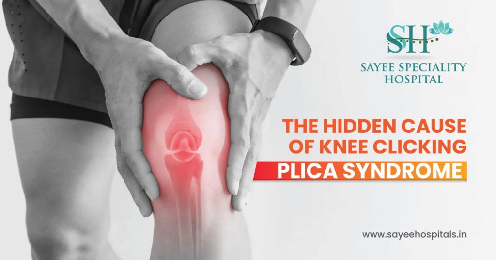
The Hidden Cause Of Knee Clicking : Plica Syndrome Knee pain is one of those things that sends most people straight down a Google rabbit hole and within about four minutes, they’ve convinced themselves they have a torn meniscus. It’s the most talked about knee injury, so it becomes the default assumption. But there’s a lesser known condition that mimics a meniscus tear closely enough to fool not just patients, but clinicians too, and it’s been quietly responsible for a lot of misdiagnosed, mistreated knee pain. It’s called plica syndrome. And once you understand what it is, that clicking, catching, aching knee of yours might start to make a lot more sense. So What Even Is a Plica? Most people have never heard the word, which is fair, it doesn’t come up much outside orthopaedic consultations. A plica is simply a fold in the synovial membrane, the thin tissue that lines the inside of your knee joint. Most knees have four of them, and in the majority of people, they’re completely harmless, present from embryonic development, just sitting there doing nothing in particular. The one that causes trouble is the medial plica, the fold that runs along the inner side of the knee. When this fold gets repeatedly irritated, through overuse, repetitive movement, or a direct knock, it thickens and tightens. Instead of gliding smoothly over the structures around it, it starts catching and dragging across the femoral condyle (the rounded end of the thigh bone) with every bend of the knee. That repeated friction inflames the tissue further, which makes it thicker, which makes the catching worse. It becomes a self perpetuating cycle. The result is medial plica syndrome. a condition that produces pain, clicking, and swelling in a pattern that looks, on the surface, almost identical to a meniscal injury. Plica Syndrome vs. Meniscus Tear :The Key Difference This is where the diagnosis gets tricky, and where a lot of people end up in the wrong treatment pathway. Both conditions produce knee pain, both can cause swelling, and both can create that unsettling sensation of instability, like the knee might give way. So it’s not hard to see why one gets mistaken for the other. The distinction that matters most is location. Meniscal pain tends to sit right at the joint line, that narrow gap between the femur and tibia that you can feel along the inner or outer edge of the knee. Plica syndrome pain typically sits above the joint line, toward the middle of the knee, often described as being just below or around the kneecap rather than at the sides. The mechanical symptoms are also slightly different in character. People with plica syndrome frequently describe a clicking or snapping sensation when bending the knee, not a vague discomfort but a distinct, repeatable mechanical event. A catching feeling when standing up after sitting for a while is another common one, that brief moment where the knee seems to stick before it moves freely. Some people can actually feel a tender, thickened band of tissue when they press just to the inner side of the kneecap, that’s the irritated plica itself. None of this is definitive on its own, which is why imaging and clinical assessment still matter. MRI can sometimes identify a thickened plica, though it isn’t always visible even when symptomatic. The diagnosis often comes together from the full clinical picture rather than any single test. Who Gets It and Why Plica syndrome shows up most often in people who do a lot of repetitive knee flexion, runners and cyclists are the classic presentation. The repeated bending motion, thousands of times over a training block, is exactly the kind of mechanical stress that irritates the medial plica over time. It’s an overuse injury at its core, which means it tends to develop gradually rather than arriving with a specific incident. That said, it can also follow direct trauma. A dashboard injury, where the knee strikes the dashboard in a car accident, is a well documented trigger. The blunt force impact causes immediate inflammation of the synovial tissue, and if the plica happens to take the brunt of that impact, the irritation and thickening can follow. Age plays a role too. Younger, active individuals make up the majority of cases, simply because they’re generating the repetitive loading that drives the condition. But it’s not exclusively a young person’s problem, anyone with a physically active lifestyle and the right anatomy can develop it. Treatment and Why Most People Don’t Need Surgery Here’s the genuinely reassuring part: plica syndrome responds well to conservative treatment in the majority of cases. Surgery is very much the last resort, not the starting point. The foundation of non-surgical management is strengthening the quadriceps. Strong quads reduce the mechanical load on the knee joint and change how forces are distributed across the structures inside it, including the plica. Hamstring stretching matters too, because tight hamstrings alter knee mechanics in ways that increase friction at the plica. A structured physiotherapy programme that addresses both is usually the most effective starting point. Rest from the aggravating activity, ice for acute inflammation, and anti-inflammatory medication to settle the irritated tissue round out the conservative approach. Many patients see significant improvement within several weeks of consistent management, particularly if they catch it early before the plica has become severely thickened. For cases that don’t respond, where the plica has become substantially thickened and fibrotic, or where symptoms have been going on long enough that conservative measures aren’t shifting them arthroscopic resection is the surgical option. It’s minimally invasive: the surgeon uses a small camera and instruments through tiny incisions to locate and remove the irritated fold. Recovery is typically around six weeks, and outcomes are generally very good when the diagnosis has been confirmed properly before going in. The Takeaway If your knee has been clicking, catching, or aching in a way that nobody has been able to fully explain and especially if you’ve already been
Cyclic Vomiting Syndrome: More Than Just A Stomach Bug

Cyclic Vomiting Syndrome: More Than Just A Stomach Bug Every parent knows the drill when a child starts vomiting. You assume it’s a bug, you set up camp on the bathroom floor, and you wait for it to pass. And it does, usually within a day or two. But for some children, and a significant number of adults, the vomiting comes back. Same intensity, same pattern, same misery weeks or months later, like clockwork. And no virus, no dodgy food, no obvious explanation. If that pattern sounds familiar, it has a name. Cyclic vomiting syndrome is a real, recognised condition and it’s still nowhere near as well understood by the general public as it should be. What Makes CVS Different From a Normal Stomach Bug The defining feature of cyclic vomiting syndrome isn’t the vomiting itself, it’s the pattern. Episodes are intense, they’re recurrent, and crucially, the person returns to completely normal health in between them. Not slightly under the weather. Not still recovering. Genuinely fine, sometimes for weeks or months at a stretch, until the next episode arrives and the whole thing starts again. That cycle severe illness, then complete wellness, then severe illness again, is what separates CVS from most other gastrointestinal conditions. A stomach bug resolves and doesn’t come back in the same predictable way. Inflammatory bowel conditions tend to produce ongoing symptoms rather than discrete episodes with clear symptom free windows. CVS has a rhythm to it that, once you recognise it, is fairly distinctive. It can start at any age, but children are disproportionately affected. Many cases that begin in childhood either resolve as the child gets older or evolve into migraine, which gives you a clue about the underlying biology. The Four Phases Because CVS Has a Structure One of the things that makes CVS manageable, at least in part, is that most patients experience episodes in a recognisable sequence. Understanding those phases helps both patients and families know what’s coming and respond appropriately at each stage. The prodrome phase is the warning window, minutes to hours before vomiting begins, there’s often a wave of nausea, sweating, and a general sense that something is about to happen. Many patients report this striking in the early hours of the morning. For some, this phase is long enough to take abortive medication and potentially head off the full episode. The vomiting phase is the acute stage intense, frequent nausea and vomiting, sometimes projectile, sometimes accompanied by retching without producing anything. Light and sound sensitivity are common during this phase, which again points toward the migraine connection. Most patients need a dark, quiet room and are essentially non functional until it passes. This phase can last anywhere from a few hours to several days, and the dehydration risk during prolonged episodes is real. The recovery phase is when vomiting and nausea gradually ease off, energy starts returning, and appetite tentatively comes back. This can feel fragile patients often describe feeling cautiously optimistic, not wanting to jinx it. And then the well phase the symptom free interval where the person genuinely feels like themselves again. This is what makes CVS so confusing to outside observers. Someone who was hospitalised and severely unwell last week looks and feels completely normal today. That contrast is hard to reconcile, and it contributes to how often CVS gets dismissed or misunderstood. What’s Actually Causing It Honest answer: not fully understood yet. But the working model describes CVS as a brain-gut disorder, a dysfunction in the communication pathways between the central nervous system and the digestive system. The gut has its own extensive nervous system, and in CVS, something in that signalling goes wrong in a recurring, episodic way. The strongest established link is with migraines. Children with CVS have a significantly higher rate of migraine headaches, and many adults who were diagnosed with CVS in childhood go on to develop classic migraines. The underlying neurovascular mechanism appears to be related, which is partly why anti migraine medications end up being part of the treatment picture. Triggers vary between individuals, but some come up repeatedly. Emotional stress is a major one and interestingly, it doesn’t have to be negative stress. Heightened excitement, anticipation, big events can trigger episodes just as reliably as anxiety or upset. Certain foods appear on many patients’ trigger lists chocolate, cheese, caffeine, and MSG are the most commonly cited. Physical illness, sleep disruption, and hormonal changes around menstruation are also documented triggers in some patients. Identifying your personal trigger pattern, if there is one, is one of the most practically useful things a CVS patient can do. Getting to a Diagnosis This is where CVS gets frustrating. There’s no single test that confirms it. Diagnosis is reached by exclusion, ruling out other conditions that could explain recurrent vomiting before landing on CVS as the explanation. That process typically involves blood tests checking metabolic markers and organ function, abdominal imaging via ultrasound or CT scan, and often endoscopy to assess the upper GI tract. In children, additional investigations may look at whether there’s a metabolic or mitochondrial component. The Rome IV diagnostic criteria provide a clinical framework broadly, recurrent stereotyped episodes of vomiting with return to baseline health in between, over a defined period but arriving at that diagnosis in practice often takes time and multiple specialist appointments. For families, that journey can be exhausting. Episodes keep happening while investigations continue, and the absence of a clear diagnosis for months or years is its own particular strain. Managing CVS What Treatment Actually Looks Like Treatment in CVS is phase specific, which means what you do during an episode is different from what you do between episodes. During the vomiting phase, the priorities are preventing dehydration and managing the nausea enough to allow some rest. Mild to moderate episodes can sometimes be managed at home with oral anti nausea medication and careful fluid intake. More severe or prolonged episodes often require hospital admission for IV fluids and stronger antiemetics. Some
MRKH Syndrome Anatomy, Diagnosis And Hope

MRKH Syndrome Anatomy, Diagnosis And Hope Getting your first period is something most teenage girls expect to happen. When it doesn’t, when 15 comes and goes, then 16, while friends around you are navigating something you haven’t experienced, the confusion and quiet anxiety that follows can be difficult to put into words. For some young women, that absence eventually leads to a diagnosis that changes how they understand their own body entirely. Mayer Rokitansky Küster Hauser syndrome – MRKH for short is one of those diagnoses. It’s rare, it’s often poorly understood outside specialist circles, and it arrives at an age when most young women are already navigating enough. Here’s what it actually means, what it doesn’t mean, and why the word “hope” in the title is genuinely earned. What MRKH Actually Is MRKH syndrome is a congenital condition meaning it’s present from birth in which the uterus, cervix, and upper portion of the vagina either fail to develop fully or are absent entirely. It affects roughly 1 in 4,500 females and is caused by incomplete development of the Müllerian ducts, the embryonic structures that would normally form these reproductive organs. That’s why it’s also called Müllerian agenesis. Here’s the part that surprises most people when they first hear it: the ovaries are completely unaffected. Women with MRKH have normal ovarian function, normal female chromosomes (46,XX), and normal external anatomy. Oestrogen is produced, puberty progresses typically, breast development, pubic hair, everything, because all of that is driven by the ovaries, not the uterus. The absence is internal and structural, not hormonal. Type 1 and Type 2 What’s the Difference MRKH is broadly categorised into two forms, and the distinction matters for how thoroughly a patient needs to be investigated at diagnosis. Type 1 – sometimes called isolated MRKH – involves the reproductive anatomy only. The uterus, cervix, and upper vagina are absent or underdeveloped, and nothing else is affected. Type 2 – often referred to as MURCS association, involves the same reproductive underdevelopment alongside abnormalities in other organ systems. The kidneys are most commonly involved, a single kidney, a kidney located in an unusual position, or a horseshoe kidney are all documented associations. Skeletal anomalies, particularly scoliosis or vertebral differences, are also seen. Hearing differences and, more rarely, cardiac abnormalities round out the picture. This is why a diagnosis of MRKH isn’t just a gynaecological workup, it typically involves renal imaging, spinal assessment, and hearing evaluation as well, to establish which type a patient has and whether any additional conditions need monitoring. How It Gets Diagnosed Most MRKH diagnoses happen in adolescence, prompted by primary amenorrhoea, the absence of a first menstrual period by around age 15 in a girl who is otherwise going through puberty normally. That combination, puberty progressing typically but no period arriving, is the classic presentation that sends clinicians investigating. The diagnostic workup usually includes pelvic ultrasound as a starting point, followed by MRI of the pelvis for more detailed anatomical mapping. Blood tests check hormone levels and confirm chromosomal profile. The physical examination and imaging together build a picture that distinguishes MRKH from other causes of primary amenorrhoea, including androgen insensitivity syndrome, which can present similarly but has a different underlying mechanism. An important detail worth noting: because the ovaries are functional, some women with MRKH experience monthly cramping and pelvic discomfort even without bleeding. The ovaries still ovulate, endometrial like tissue can still respond cyclically, and the resulting pain, with no outlet, can be genuinely significant. This symptom is sometimes dismissed or goes unexplained for months before a diagnosis is made. Treatment and Management There’s no single treatment for MRKH, management is tailored around each person’s anatomy, priorities, and wellbeing. For vaginal lengthening, progressive dilation therapy is the standard first-line approach. Using graduated dilators over a period of months, most women achieve a functional vaginal canal without surgery. It requires consistency and patience, but success rates with proper guidance are high and the approach avoids surgical risk entirely. For women who are unable to complete dilation or prefer a surgical route, procedures using tissue grafts – the Vecchietti or McIndoe techniques among others, are available options. General healthcare follows some modified pathways. Cervical cancer screening isn’t applicable in the absence of a cervix, but HPV vaccination is still recommended since vaginal and vulval HPV related conditions remain relevant. STI screening and sexual health care continue as normal. Cyclic pelvic pain, where present, is typically managed with ibuprofen or hormonal medication to suppress the cycle. The psychological dimension of an MRKH diagnosis deserves equal weight to the physical. Being told at 15 or 16 that your reproductive anatomy is significantly different from what you expected and processing what that means for your identity, relationships, and future, is not a small thing. Specialist psychosocial support, peer support networks, and counselling are all recognised parts of comprehensive MRKH care, not optional extras. Fertility and the Path to Parenthood This is often the question that sits heaviest after a diagnosis, particularly for young women who haven’t yet had the chance to think about whether or not they want children. Carrying a pregnancy isn’t possible without a uterus. That’s the straightforward reality. But biological motherhood, in the sense of having children who are genetically your own, is a genuine option, because the ovaries function normally. Eggs can be retrieved through IVF stimulation and fertilised, and the resulting embryo carried by a gestational surrogate. Uterine transplantation is also an emerging possibility, with successful pregnancies following transplant now documented in several countries, though it remains experimental and not yet widely available. For women who choose not to pursue biological parenthood, adoption and fostering are paths that many in the MRKH community have navigated with tremendous fulfilment. None of these paths are without complexity. But options exist, more than existed even a decade ago and that matters. A Final Word An MRKH diagnosis changes things. It changes how a young woman understands her body, it raises questions she wasn’t expecting to
A Guide To Choosing Heat OR Cold Therapy

A Guide To Choosing Heat OR Cold Therapy It’s one of those decisions that sounds simple until you’re actually standing in front of the freezer at midnight, ankle swollen, wondering whether to grab the ice pack or dig out the heating pad. Most people just guess. And honestly, guessing wrong doesn’t ruin anything, but getting it right genuinely speeds up recovery, sometimes by a meaningful amount. So here’s a straightforward breakdown of how both actually work, when to use each one, and what to avoid. Heat Therapy What It Does and When It Helps Heat works by drawing more blood to an area. Warmth causes blood vessels to dilate, circulation increases, and muscles, which are essentially protein fibres that respond directly to temperature begin to relax and lengthen. Tissue becomes more pliable, stiffness eases, and that deep aching tension that builds up in overworked muscles starts to loosen its grip. That’s why heat is the right choice for chronic pain, muscle tightness, and joint stiffness, the kind of discomfort that’s been sitting there for days, not hours. A stiff lower back on a cold morning, persistent neck tension from desk work, achy joints flaring up in winter. These are heat situations. There are two ways to apply it, dry heat, like electric heating pads or heat patches, and moist heat, like a warm damp towel, a hot bath, or a steam pack. Moist heat tends to penetrate deeper into the tissue in less time, which is why a long hot bath often feels more effective than a heating pad on a sore back. For most applications, 15 to 20 minutes is enough. Warm, not hot, if it’s uncomfortable to hold against your skin, it’s too hot. The one firm rule: never apply heat to an acute injury. Fresh swelling, bruising, or a recently injured area still generating inflammation, heat makes all of that worse. It increases blood flow to a site that’s already overloaded with it. Cold Therapy What It Does and When It Helps Cold does the opposite. It constricts blood vessels, reduces blood flow to the area, and slows the inflammatory response. That’s exactly what you want in the first 24 to 72 hours after an acute injury a sprained ankle, a pulled muscle, a knocked knee. The goal in those early hours isn’t comfort so much as damage control. Limiting the extent of swelling and inflammation in that initial window affects how quickly the tissue heals afterward. Cold also numbs the nerve endings in the treated area, which gives fairly immediate short-term pain relief. That’s the secondary benefit, the primary one is managing the inflammatory response while it’s still acute. At home, always put something between the ice pack and your skin, a folded tea towel works fine. Direct ice to skin contact, especially for longer than a few minutes, can cause ice burns and superficial tissue damage. Keep applications to 10 to 20 minutes, and repeat several times across the day rather than leaving it on continuously. Where cold goes wrong: applying it to stiff muscles or chronic joint problems. Cold tightens tissue. On a muscle that’s already tight, that just makes things worse and can increase the risk of a strain if you then try to move. The Easy Rule to Remember If it’s new, it’s swollen, it just happened use cold. If it’s been there a while, it’s stiff, it aches without swelling, use heat. And if you’re genuinely unsure whether an injury is acute or chronic, default to cold in the first 48 hours, then reassess. Who Should Be Careful Both therapies are generally safe for healthy adults, but a few groups need to think more carefully before using them. People with diabetes, peripheral vascular disease, or any condition that affects nerve sensation need to be cautious with both. Reduced sensation means you may not feel the warning signs of skin damage, burning or frostbite, until it’s already happened. If there’s any numbness or circulation issues in the area you’re treating, check with your doctor first. Anyone with a heart condition or high blood pressure should get professional guidance before using saunas, hot baths, or cold immersion. Sudden temperature extremes place real demand on the cardiovascular system, vasodilation from heat and vasoconstriction from cold both affect how hard the heart has to work. And practically speaking: if you’re using cold therapy and the injury isn’t improving after 48 hours, that’s a prompt to seek proper assessment. If heat has been your go to for a pain that’s persisted beyond a week without improvement, that’s also worth getting looked at. Both therapies manage symptoms, they don’t fix underlying problems. Recent Posts A Guide To Choosing Heat OR Cold Therapy A Guide To Choosing Heat OR Cold Therapy It’s one of those decisions that sounds… The Pressure Trap: Understanding Compartment syndrome The Pressure Trap: Understanding Compartment syndrome Most people have never heard of compartment syndrome until… The Barking Cough: A Parent’s Guide To Croup The Barking Cough: A Parent’s Guide To Croup There’s a specific kind of panic that… The Silent Emergency: Understanding Ovarian Torsion The Silent Emergency: Understanding Ovarian Torsion Most people have never heard of ovarian torsion. And… edit post A Guide To Choosing Heat OR Cold Therapy A Guide To Choosing Heat OR Cold Therapy… edit post The Pressure Trap: Understanding Compartment syndrome The Pressure Trap: Understanding Compartment syndrome Most people… edit post The Barking Cough: A Parent’s Guide To Croup The Barking Cough: A Parent’s Guide To Croup… edit post Influenza In Children: Causes, Symptoms, And Prevention Influenza In Children: Causes, Symptoms, And Prevention Influenza,…
The Pressure Trap: Understanding Compartment syndrome

The Pressure Trap: Understanding Compartment syndrome Most people have never heard of compartment syndrome until it happens to them or someone they love. And by the time it does, there usually isn’t much time to start researching. That’s the problem with this condition. It moves fast, it gets mistaken for something less serious, and the consequences of missing it can be permanent. So here’s what you need to understand before you ever need to use it. Your Muscles Live in Compartments and That Matters Wrapped around every muscle group in your body is a layer of dense, fibrous tissue called fascia. It organises muscles, nerves, and blood vessels into tightly contained sections compartments and unlike skin or fat, fascia doesn’t stretch. It doesn’t give. It holds its shape regardless of what’s happening inside it. That’s normally fine. Under normal circumstances, the pressure inside these compartments stays at a level that allows blood to flow freely, nerves to function properly, and muscle tissue to stay healthy. But when something causes pressure to build inside one of these sealed spaces bleeding, swelling, fluid accumulation the fascia doesn’t accommodate it. The pressure has nowhere to go. And as it climbs, it starts compressing the blood vessels and nerves running through that compartment. Cut off the blood supply long enough, and the tissue inside starts to die. That’s compartment syndrome in its simplest form, a pressure problem with a rapidly closing window. Acute VS Chronic: Two Very Different Situations This is worth clarifying upfront, because the two types of compartment syndrome are almost nothing alike in terms of urgency. Acute compartment syndrome is a surgical emergency. Full stop. It typically follows a significant traumatic injury, a serious fracture (the tibia, or shin bone, is one of the most common triggers), a crush injury, a severe fall, or a high impact accident. The injury causes internal bleeding or swelling within the compartment, pressure spikes rapidly, and the clock starts ticking. Without a fasciotomy the surgical procedure to relieve that pressure permanent muscle and nerve damage can occur within hours. In the worst cases, the consequences include paralysis, extensive tissue necrosis, kidney failure from the breakdown products of dying muscle, or death. Chronic exertional compartment syndrome is a completely different beast. It’s not life threatening, it’s not a sudden emergency, and it primarily affects athletes particularly runners, cyclists, and anyone doing high volume repetitive leg training. During intense exercise, muscle volume increases as blood floods into the tissue. Normally, the fascia accommodates this to a degree. In people with chronic compartment syndrome, it doesn’t pressure builds during activity, causes pain and tightness, and then eases again once they stop. It’s a pattern. It’s predictable. And for competitive athletes, it can be genuinely career derailing even though it won’t kill them. Both conditions are real and deserve attention, they just require very different levels of urgency. What It Actually Feels Like With acute compartment syndrome, the phrase clinicians use most often is “pain out of proportion.” A broken bone hurts that’s expected. But compartment syndrome produces a deep, relentless, escalating ache that doesn’t respond to normal pain relief and keeps getting worse rather than plateauing. Patients often describe it as a feeling that the muscle is going to burst through the skin. Alongside that pain, look for a sensation of extreme tightness or fullness in the affected limb, like the tissue is being squeezed from the inside. You might notice visible firmness or slight bulging over the compartment. Paresthesia pins and needles, numbness, or a strange tingling, indicates that nerve compression is already underway. Pain that intensifies when the muscles in that compartment are passively stretched is another hallmark sign that clinicians specifically test for. In the chronic version, the pattern is different. Pain and tightness usually come on at a predictable point during exercise often after a certain distance or duration and ease fairly quickly once activity stops. There’s less of the severe aching, more of a burning, cramping tightness. Paresthesia can occur here too, usually in the foot or lower leg. Getting a Diagnosis For acute cases in a trauma or emergency setting, diagnosis is often clinical, a combination of the injury mechanism, symptom pattern, and physical examination findings. But when there’s genuine uncertainty, compartment pressure measurement confirms it. A specialised needle is inserted directly into the muscle compartment to obtain a real time pressure reading. A normal resting compartment pressure sits below around 15 mmHg. Readings approaching or exceeding 30 mmHg particularly when the gap between that pressure and the patient’s diastolic blood pressure narrows indicate that fasciotomy is needed immediately. For chronic exertional compartment syndrome, pressure readings are taken both before and after exercise to capture how the numbers shift with activity. This before and after comparison is usually what clinches the diagnosis, since resting pressures in these patients are often normal. Treatment: From Surgery to Rehab Acute compartment syndrome has one treatment: fasciotomy. The surgeon makes incisions through both the skin and the fascia, releasing the pressure and restoring blood flow to the trapped tissue. It’s not a subtle procedure, the wounds are left open initially and closed in a second surgery once the swelling has reduced, but it works. Done in time, it saves limbs and lives. Done too late, or not at all, the outcomes are significantly worse. Chronic compartment syndrome, by contrast, rarely requires surgery as a first step. Most patients see meaningful improvement through a combination of activity modification, biomechanical assessment, custom orthotics, and a structured physical therapy programme that addresses running gait or training load. Anti-inflammatory medications can help manage symptoms during the adjustment period. For athletes who don’t improve with conservative management and want to return to full competitive training, a fasciotomy can be performed electively and results are generally very good. Why This Condition Gets Missed Compartment syndrome gets missed for a straightforward reason: the pain gets attributed to the injury that caused it. Of course a broken leg hurts. Of course a crush
The Barking Cough: A Parent’s Guide To Croup

The Barking Cough: A Parent’s Guide To Croup There’s a specific kind of panic that sets in at 2am when your toddler wakes up making a sound you’ve never heard before, a harsh, seal like bark that doesn’t sound anything like a normal cough. If you’ve been through it, you know exactly what I’m describing. If you haven’t, just know that the first time you hear it, your instinct is to grab your keys and head straight to A&E. Sometimes that’s the right call. But more often than not, what you’re dealing with is croup, a common childhood respiratory illness that sounds far scarier than it usually is. Here’s what you actually need to know. So What Is Croup, Exactly? Croup isn’t a single disease, it’s a pattern of symptoms triggered most commonly by the parainfluenza virus, though other respiratory viruses can cause it too. The virus infects the upper airway, leading to inflammation and swelling around the larynx (the voice box), the trachea (the windpipe), and the bronchi further down. That swelling is what produces the distinctive barking cough and, in more significant cases, a condition called stridor, a high pitched, strained sound when the child breathes in. The reason young children are so much more affected than older kids or adults comes down to anatomy. A toddler’s airway is narrow to begin with, genuinely quite small in diameter. So when inflammation reduces that space even slightly, the effect on breathing is disproportionately large. The same degree of swelling that would give an adult a scratchy throat can meaningfully restrict a two year old’s airway. As children grow and their airways widen, croup becomes less of a concern it’s rare in children over six, and almost unheard of in older kids. How It Usually Starts and What to Watch For: Croup has a habit of announcing itself as something completely ordinary. It usually begins with a day or two of cold-like symptoms runny nose, mild fever, a bit of a sore throat. Nothing alarming. Then, often overnight, the cough changes. It becomes loud, harsh, and unmistakably barky. Some parents describe it as a dog barking; others say a seal. Either way, once you’ve heard it, you won’t mistake it for anything else. Along with the cough, your child may develop a hoarse voice again, because the larynx is swollen and vibrating differently. Stridor, that high pitched inhale sound, tends to appear when symptoms are more significant and the airway is more noticeably narrowed. One thing worth knowing: croup almost always gets worse at night. The exact reason isn’t fully understood, but changes in airway tone during sleep, combined with the horizontal position, seem to play a role. Children who seemed manageable at bedtime can sound considerably worse by midnight, which is a big part of why croup ends up being such a common reason for late-night emergency visits. Crying and distress also worsen things. An upset child breathes faster and harder, which irritates an already inflamed airway and the worse they breathe, the more frightened they get. It becomes a cycle that can escalate quickly if you can’t get them settled. Managing Croup at Home: Because croup is viral, antibiotics won’t touch it. Most mild-to-moderate cases can be managed at home, and the focus is really on two things: keeping your child calm and keeping them comfortable. Calm is genuinely the most important thing. A settled child breathes slower and easier, which breaks the distress cycle. Hold them upright, either sitting up or with their head elevated as lying flat can make the breathing feel worse. Sit with them, talk quietly, read a story, put on something familiar on a screen. Whatever usually soothes your child, lean into it hard. Keep fluids going throughout the day. A well-hydrated airway handles inflammation better, and staying hydrated also helps loosen any mucus making things more congested. Cool, moist air is often suggested as a comfort measure ,some parents swear by sitting in a steamy bathroom for a few minutes, though the evidence on whether it genuinely helps airway symptoms is mixed. It probably doesn’t hurt, and the quiet time together may help calm an anxious child anyway. Symptoms usually peak around day two or three and then gradually improve. Most children are significantly better within five days. When Home Care Isn’t Enough: This is the part that matters most, so pay attention here. Most croup is mild. But a small percentage of cases involve significant airway obstruction, and those need medical treatment, typically an oral or inhaled corticosteroid to reduce inflammation, or in severe cases, nebulised epinephrine in a hospital setting. Call 108 or go straight to your nearest emergency department if: Your child is producing stridor while sitting still and calm not just when crying or coughing, but at rest. That’s a sign of meaningful airway narrowing. Their breathing is rapid and visibly laboured. If you can see the muscles between their ribs or at the base of their throat pulling inward with each breath, their body is working hard to move air. There’s any bluish or greyish discolouration around the lips, nose, or fingertips. This is called cyanosis it means oxygen levels are dropping and this is a genuine emergency. They’re drooling excessively or struggling to swallow. This is less typical of straightforward croup and can suggest a different, more serious condition affecting the airway that needs urgent assessment. If something just feels wrong, if your instincts are telling you your child is in real difficulty trust that. Parents are often right. Medical staff would always rather assess a child who turns out to be fine than have a family delay seeking help. The Bigger Picture: Croup is one of those conditions where knowledge genuinely makes a difference. A parent who recognises that barking cough for what it is, knows how to keep their child calm, and understands exactly which symptoms require emergency care is in a much stronger position than one caught completely off guard at midnight.
The Silent Emergency: Understanding Ovarian Torsion
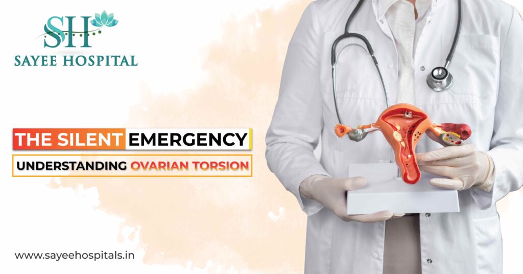
The Silent Emergency: Understanding Ovarian Torsion Most people have never heard of ovarian torsion. And that’s a problem, because when it happens, you don’t get the luxury of time to Google your symptoms, call your mum, or wait until Monday to see your GP. This is the kind of emergency that unfolds in hours, not days, and the difference between keeping your ovary and losing it often comes down to a single decision: going to A&E now, or convincing yourself it’ll pass. So let’s talk about it properly. What’s Actually Happening Inside the Body Your ovaries aren’t free floating, they’re held in position by a set of ligaments that keep everything anchored within the pelvic cavity. Think of it like a suspension system. When ovarian torsion occurs, one ovary rotates around those ligaments, essentially twisting itself. That twist clamps down on the blood vessels feeding the ovary, the arteries and veins that keep the tissue alive. Once blood flow is cut off, you’re on a clock. The ovarian tissue starts to starve. Without oxygenated blood reaching it, ischemia sets in, and if the torsion isn’t surgically corrected in time, that tissue dies. A process called necrosis. At that point, the ovary typically can’t be saved. Surgeons estimate the window between symptom onset and permanent organ loss can be as short as six hours in severe cases. Sometimes less. That’s not a lot of time to second guess yourself. The Pain and Why It’s So Easy to Dismiss The most recognisable sign is sudden, one sided pelvic pain. Not gradual. Not a slow build. It hits fast, and when women describe it, they consistently reach for words like “stabbing,” “sharp,” or simply “the worst pain I’ve ever felt.” It usually settles on whichever side the affected ovary is on, but it can radiate into the lower back, along the flank, sometimes down toward the inner thigh. What catches a lot of people off guard is something called intermittent torsion, or torsion detorsion. The ovary twists, then untwists on its own. The pain eases. And naturally, you think whatever was wrong has sorted itself out. But that’s not what’s happening each episode is causing further damage, and the risk of a full, irreversible torsion climbs with each cycle. Feeling better for a few hours doesn’t mean you’re out of danger. Alongside the pain, most women also experience nausea and vomiting often severe enough to be mistaken for food poisoning or a stomach bug. Fever and abnormal bleeding can appear in later stages, once tissue breakdown has already begun. If you’re ticking more than one of these boxes at once, please don’t wait it out at home. Who Gets Ovarian Torsion and Why Technically, it can happen at any age. There are documented cases in young girls and in postmenopausal women. But the large majority of cases occur in women between roughly 20 and 40 the reproductive years and there are a handful of factors that meaningfully raise the risk. The biggest one is an ovarian cyst or mass. When a cyst grows beyond about 5 centimetres, it throws off the balance of the ovary. That extra, asymmetric weight makes the ovary unstable more likely to flip, more likely to twist. Dermoid cysts come up frequently in torsion cases, partly because they tend to grow large and are denser than other cyst types. Pregnancy creates its own particular risk. The hormonal shifts of early pregnancy loosen the ligaments that normally keep the ovary stable, while corpus luteum cysts, which are a normal part of early pregnancy, add the kind of weight that tips things toward torsion. It’s a frustrating combination of factors that come together at exactly the wrong time. Women going through IVF or other ovarian stimulation protocols are also in a higher risk category. When the ovaries are stimulated to produce multiple follicles, they can enlarge significantly. Ovarian hyperstimulation syndrome, or OHSS, is a well-recognised complication of fertility treatment and enlarged, hyperstimulated ovaries are considerably more prone to torsion. If you’re mid-cycle on a stimulation protocol, knowing the warning signs isn’t optional. One more thing worth noting: if you’ve had ovarian torsion before, your remaining ovary carries an elevated risk. It’s not a guarantee, but it’s a reason to stay alert. Getting a Diagnosis and Why Imaging Isn’t Always Enough When torsion is suspected in an emergency setting, a transvaginal ultrasound with Doppler imaging is usually the starting point. Doppler lets clinicians assess blood flow to the ovary, and absent or reduced flow is a strong indicator of torsion. But here’s the catch: normal blood flow on Doppler doesn’t rule it out. Torsion detorsion cases can show intermittent, preserved flow and still be causing serious damage. Imaging alone has its limits. MRI can offer more detail the “whirlpool sign,” a twisted vascular pedicle, is a telling feature, but in an acute emergency, there often isn’t time for it. Diagnostic laparoscopy remains the definitive answer. A small incision, a camera, and the surgeon can see exactly what’s happening. More importantly, they can act on it immediately. If torsion is confirmed, the ovary is manually untwisted, detorsion, to restore circulation. Even if the ovary looks discoloured or dusky at the time of surgery, most gynaecologists will still attempt to save it. Appearances can be deceiving; many ovaries that looked non-viable in theatre have gone on to recover fully. If a cyst caused the problem, it’s typically removed in the same procedure. In recurrent cases, oophoropexy, surgically anchoring the ovary may be considered to reduce the chance of it happening again. The Honest Truth About Prevention There’s no way to prevent ovarian torsion entirely. No pill, no diet, no exercise routine changes the underlying mechanics. What you can control is your response to symptoms. Women with known cysts should have a conversation with their gynaecologist about monitoring frequency and what to watch for. Women undergoing fertility treatment deserve a clear briefing on torsion symptoms before stimulation begins, not buried in a leaflet, but
Influenza In Children: Causes, Symptoms, And Prevention

Influenza In Children: Causes, Symptoms, And Prevention Influenza, commonly known as the flu, is a contagious viral infection that affects the respiratory system, particularly the lungs and airways. While most children recover within a week, some may develop severe complications requiring hospitalization. In extreme cases, the flu can lead to pneumonia or even death. Types of Influenza Viruses Influenza A & B: These strains cause seasonal outbreaks, particularly in winter, leading to widespread infections and increased hospitalizations. Since these viruses mutate frequently, new strains emerge yearly. Influenza C: Unlike A and B, this strain is less common, does not follow a seasonal pattern, and causes mild respiratory symptoms. How Influenza Spreads Children contract the flu virus through: Respiratory droplets released when an infected person coughs or sneezes. Direct contact with contaminated saliva or surfaces. Sharing items like toys, stationery, and electronic devices with infected individuals. Flu viruses can spread even before symptoms appear and remain contagious for up to a week after infection. Common Symptoms Children with influenza may experience: High fever (103–105°F or 39.4–40.5°C) Muscle and joint pain Runny or blocked nose Persistent cough Nausea and vomiting Fatigue and weakness Headache and sore throat Diarrhea (in some cases) Diagnosis of Influenza Doctors diagnose the flu based on symptoms, but laboratory tests like Polymerase Chain Reaction (PCR) can confirm the presence and strain of the virus. PCR tests are highly sensitive and help in detecting influenza more accurately than traditional methods. Preventing Influenza in Children To reduce the risk of flu infections: Annual flu vaccinations are essential, especially for children under five and those with weak immune systems. Hygiene practices: Teach children to wash hands frequently and cover their mouth while coughing or sneezing. Avoiding exposure: Keep children away from infected individuals. Disinfection: Regularly clean shared items like toys, towels, and school supplies. Treatment and Home Care Ensure children stay hydrated and get ample rest. If symptoms worsen, consult a doctor for antiviral medications. These can reduce the severity and duration of the illness. Monitor for complications like difficulty breathing, persistent fever, or severe weakness. By prioritizing timely vaccination and proper hygiene, the risk of influenza in children can be significantly reduced. Recent Posts edit post A Guide To Choosing Heat OR Cold Therapy A Guide To Choosing Heat OR Cold Therapy… edit post The Pressure Trap: Understanding Compartment syndrome The Pressure Trap: Understanding Compartment syndrome Most people… edit post The Barking Cough: A Parent’s Guide To Croup The Barking Cough: A Parent’s Guide To Croup… edit post The Silent Emergency: Understanding Ovarian Torsion The Silent Emergency: Understanding Ovarian Torsion Most people…