


Lower Back Pain: Causes, Diagnosis, and Treatment
Lower Back Pain Causes, Diagnosis, and Treatment Almost...
Multispecialty Hospital in Padappai | Sayee Specialty Hospital
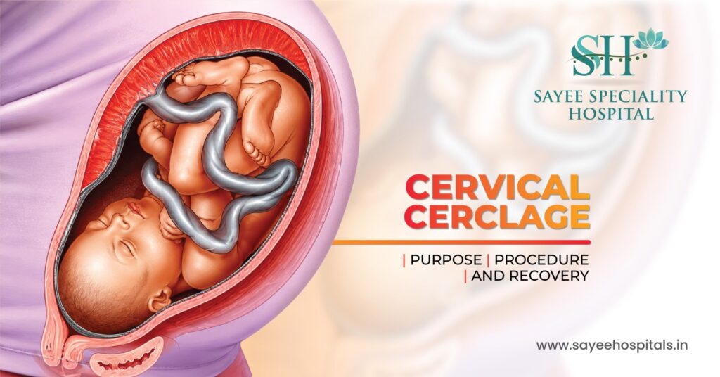
Cervical Cerclage: Purpose,Procedure & Recovery
Most people go through pregnancy without ever hearing the term cervical cerclage. For those who do encounter it, it usually arrives at a moment of heightened anxiety, a scan that shows concerning cervical shortening, a history of second-trimester loss that nobody fully explained at the time, or a conversation with a specialist that suddenly reframes the entire pregnancy as higher risk than expected.
Understanding what a cerclage actually is, why it’s recommended, and what the procedure involves can take a significant amount of fear out of that experience. It’s a well-established intervention with a clear purpose and a straightforward recovery, and for the pregnancies where it’s indicated, it makes a genuine difference.
The cervix is the lower portion of the uterus, a cylindrical structure, typically firm and closed during pregnancy, that keeps the uterine contents secure until the body is ready for labour. In a healthy pregnancy, the cervix remains long, closed, and firm throughout the second and into the third trimester, only beginning to soften, shorten, and dilate as labour approaches.
In some pregnancies, the cervix begins to weaken and shorten prematurely, a condition referred to clinically as cervical insufficiency or an incompetent cervix. The cervical tissue loses its structural integrity earlier than it should, and without intervention, this can lead to the cervix opening silently and painlessly during the second trimester, resulting in pregnancy loss or extremely premature delivery.
Several factors can contribute to cervical insufficiency. Previous cervical procedures LEEP (loop electrosurgical excision procedure) or cone biopsy for abnormal cervical cells, can reduce the amount of cervical tissue available and affect structural integrity. Previous significant cervical dilation during surgical procedures, uterine abnormalities, connective tissue disorders, and a history of second-trimester losses all raise the index of suspicion. In some cases, cervical shortening is identified incidentally on a routine transvaginal ultrasound during the second trimester, before any symptoms have appeared.
Cerclage isn’t recommended for every high-risk pregnancy, it’s specifically indicated for cervical insufficiency, and the decision is based on a combination of obstetric history and ultrasound findings.
A history-indicated cerclage is placed based on previous pregnancy losses or preterm births consistent with cervical insufficiency, typically planned and placed between weeks 12 and 14 before any cervical change is visible. An ultrasound-indicated cerclage is placed when transvaginal ultrasound during the current pregnancy shows cervical shortening below a certain threshold typically a cervical length below 25mm in a woman with a prior preterm birth. An emergency or rescue cerclage is placed when the cervix is already dilating in the second trimester, which is a more urgent situation with different risks and outcomes than planned placement.
The decision involves careful risk-benefit assessment cerclage isn’t without risk, and it’s not beneficial in all situations where preterm birth is a concern. Preterm labour driven by infection or contractions, for instance, is not improved by cerclage and may be worsened by it. The specialist’s judgement about the specific mechanism driving the risk matters enormously.
For most women, the anticipation of the procedure is considerably more stressful than the procedure itself. Cervical cerclage is typically performed as an outpatient procedure, meaning the same day return home, usually under spinal or regional anaesthesia rather than general anaesthesia.
In the most common approach, the transvaginal McDonald cerclage, the patient is positioned for a standard pelvic examination. The cervix is visualised through a speculum, and strong non-absorbable sutures are placed circumferentially around the cervix, essentially drawing it closed. The procedure takes around 20 to 30 minutes in most cases. A second approach, the Shirodkar cerclage, involves a slightly more involved dissection to place the suture higher on the cervix and is used in certain clinical situations where the McDonald technique isn’t adequate.
In rare cases typically where previous transvaginal cerclages have failed, or where the cervix is too short to access vaginally, an abdominal cerclage is performed, either open or laparoscopically, placing the suture at the cervicoisthmic junction above the vaginal portion. This is a more significant procedure and is typically placed before pregnancy or in very early pregnancy.
Before the procedure, a transvaginal ultrasound confirms cervical status and foetal wellbeing. Antibiotics are commonly administered to reduce infection risk. Women are usually advised to avoid intercourse for a week before a planned cerclage.
Mild cramping and light spotting in the days immediately following the procedure are expected and don’t indicate a problem. Paracetamol is usually sufficient for discomfort management during this period. Most women are monitored for a few hours after the procedure before going home.
The recovery window of roughly 7 to 10 days requires some specific restrictions, avoiding strenuous physical activity, heavy lifting, and sexual intercourse while the cervix settles. Beyond that initial period, most women with a cerclage in place return to relatively normal daily activity, though specific guidance varies depending on the clinical situation and the individual obstetrician’s protocol.
Regular cervical length monitoring by transvaginal ultrasound continues throughout the pregnancy, typically every two to four weeks, to ensure the cerclage is holding and the cervix isn’t shortening further. Any symptoms of preterm labour, regular contractions, pelvic pressure, increased vaginal discharge, or fluid leakage, warrant immediate assessment rather than watchful waiting at home.
Cerclage is a well-established and generally safe procedure, but it isn’t without risk, and those risks are worth understanding clearly.
The most significant potential complication is premature rupture of membranes, where the suture provokes the membranes to break before the pregnancy reaches a viable or term gestation. Infection of the cervix or uterine cavity is another potential complication, which is why antibiotic prophylaxis and monitoring for signs of infection are standard parts of cerclage management. Cervical laceration, tearing at the suture site can occur, particularly if labour begins before the suture is removed.
These complications are uncommon, and the risk-benefit calculation for women with a genuine indication for cerclage overwhelmingly favours placement. But the procedure should be performed by an experienced obstetrician, and the decision should be made after thorough discussion of the specific clinical picture rather than as a precautionary measure in all high-risk pregnancies.
The cerclage sutures remain in place until around 36 to 37 weeks of pregnancy at or approaching full term. Removal is a straightforward clinic procedure requiring no anaesthesia in most cases. The sutures are cut and removed, and the cervix is then free to begin the natural process of effacement and dilation in preparation for labour.
After cerclage removal, some women go into labour relatively quickly. Others continue to term and beyond. The removal doesn’t trigger immediate labour in most cases, though it’s not uncommon for cervical changes to begin progressing more rapidly in the days following.
Women who have had an abdominal cerclage require caesarean section delivery, since the suture is placed too high to be removed vaginally that’s an important consideration in the planning of any subsequent pregnancies.
A cerclage doesn’t guarantee a term pregnancy, no intervention in obstetrics comes with guarantees. But for women with genuine cervical insufficiency, it meaningfully improves the probability of carrying the pregnancy to a viable or term gestation. That improvement in outcome is the reason the procedure exists and the reason it remains a standard part of high-risk obstetric care.
If you’ve been recommended a cerclage, the conversation with your obstetrician should cover why it’s indicated in your specific case, which technique is planned, what the recovery involves, and what monitoring will look like for the rest of the pregnancy. Going into that procedure informed and clear on the reasoning makes the experience considerably more manageable.
Recent Posts
Lower Back Pain Causes, Diagnosis, and Treatment Almost...

Strep Throat: Symptoms, Diagnosis, and Treatment Sore throats...

Understanding Menorrhagia: Causes, Diagnosis, and Treatment Most women...

Your Posture Is Destroying Your Spine Before 40...