

What a Gastroenterologist Looks for When Your Stomach Pain Won’t Go Away

What a Gastroenterologist Looks for When Your Stomach Pain Won’t Go Away Stomach pain is one of those symptoms that almost everyone has experienced, and almost no one takes seriously enough when it keeps coming back. There’s a reason for that. A single episode of abdominal discomfort rarely amounts to much – something you ate, a passing virus, a stressful week. The body sorts it out and moves on. But the stomach pain that doesn’t go away – the kind that has been there for weeks, or that disappears for a few days and reliably returns, or that gets progressively worse over months – is a different conversation entirely. That kind of pain is communicating something, and the question is whether anyone is listening carefully enough to work out what. A gastroenterologist approaching a patient with persistent abdominal pain isn’t just asking ‘where does it hurt?’. The clinical thought process is considerably more structured than that – and the pattern of details that emerge from a careful history will often point toward a diagnosis before a single test has been ordered. This post walks through how that process actually works, and what it means for someone who has been managing stomach pain on their own for longer than they should have. The History Tells More Than Most Patients Realise In gastroenterology, the clinical history is the most powerful diagnostic tool available. Imaging and endoscopy confirm – they rarely surprise. The diagnosis is usually already forming in the gastroenterologist’s mind by the time the patient finishes describing their symptoms in full. Here is what a specialist is specifically listening for: Location of the pain The abdomen is not a single compartment. Pain in the upper centre of the abdomen – the epigastric region – implicates the stomach, duodenum, pancreas, or lower oesophagus. Pain in the upper right points toward the liver, gallbladder, or bile ducts. Upper left pain is less common and can involve the spleen or splenic flexure of the colon. Pain around the navel often involves the small intestine. Lower abdominal pain – either central, right, or left – pulls the colon, appendix (on the right), ovaries, or bladder into consideration. Pain that is diffuse – spread across the whole abdomen without a consistent location – is its own diagnostic category. It can indicate a functional condition like IBS, a motility disorder, or something more generalised. The location alone narrows the differential considerably before a single question about character or timing is asked. Character of the pain – what it actually feels like Cramping or colicky pain that comes in waves and then eases points toward a hollow organ – the bowel or gallbladder – contracting against an obstruction or trying to push something through. A steady, burning sensation in the upper abdomen that is worse on an empty stomach and eased by eating suggests a duodenal ulcer. The same burning that is worse after eating and comes with regurgitation suggests gastro-oesophageal reflux or gastritis. Severe, constant pain that bores through to the back is one of the classic presentations of pancreatitis or pancreatic disease. Pain that is sharp and knife-like, particularly if it radiates, is different from pain that is dull and pressure-like, which is different again from pain that is burning. These distinctions are not academic – they directly inform the diagnostic thinking. Timing and triggers When does the pain occur? Pain that consistently comes 30 to 60 minutes after eating is different from pain that comes on an empty stomach. Pain that wakes someone from sleep at 2am has different implications from pain that never bothers them at night. Pain that is reliably worse during periods of stress points toward a functional component. Pain that has been gradually and consistently worsening over weeks – without ever fully resolving – is more concerning than pain that flares and settles. Dietary triggers tell their own story. Pain consistently after fatty meals implicates the gallbladder. Pain after gluten-containing foods that also comes with bloating and changes in stool is a flag for coeliac disease. Pain that worsens with certain vegetables and legumes has fermentation-related causes. The pattern across meals gives the gastroenterologist a picture that a blood test cannot. Associated symptoms Stomach pain doesn’t exist in isolation. A gastroenterologist will systematically ask about nausea, vomiting, changes in bowel habits, blood in the stool, unexplained weight loss, loss of appetite, fever, jaundice, and difficulty swallowing. Each accompanying symptom either broadens or narrows the differential. Abdominal pain with jaundice points to the liver or bile ducts. Pain with significant unintentional weight loss is a red flag combination that always warrants urgent investigation. Pain with alternating constipation and diarrhoea in a young adult without red flag features strongly suggests a functional cause. The Red Flag Symptoms That Change Everything Within the broader picture of chronic stomach pain, certain symptom combinations significantly raise the clinical urgency and prompt faster, more invasive investigation. These are the features a gastroenterologist is specifically watching for: Unintentional weight loss alongside abdominal pain – particularly when the weight loss is more than 5 percent of body weight over three to six months without a dietary reason Blood in the stool – whether bright red (lower gastrointestinal source) or dark and tarry (upper GI source, indicating digested blood) Vomiting blood or material that looks like coffee grounds Persistent pain that wakes the patient from sleep and doesn’t respond to standard analgesia New abdominal pain in a patient over 50 who has no prior history of GI problems Jaundice – yellowing of the skin or eyes – in conjunction with abdominal pain A palpable abdominal mass on self-examination or reported during clinical examination Family history of colorectal cancer, gastric cancer, or inflammatory bowel disease Iron deficiency anaemia without an obvious nutritional cause – chronic GI blood loss is a common and frequently missed underlying cause None of these individually confirms a serious diagnosis. But each one shifts the probability distribution enough that investigation cannot wait. A gastroenterologist seeing these
When Does a Joint Pain Actually Need Surgery? The Answer Will Surprise You

When Does a Joint Pain Actually Need Surgery? The Answer Will Surprise You Most people who walk into an orthopaedic clinic with joint pain are quietly terrified of one thing. Not the diagnosis. Not the prognosis. The possibility that someone is going to tell them they need surgery. And so they wait. Sometimes for years. They manage the pain with anti-inflammatories, limit the activities that trigger it, and avoid the orthopaedic appointment altogether because they’d rather not hear something they’re not ready for. By the time they finally come in, they’ve often been living with significantly limited function for far longer than was necessary – and occasionally, they’ve waited past the point where conservative treatment would have been most effective. Here’s what orthopaedic surgeons actually think about joint surgery that most patients don’t get told upfront: the majority of people who come in with joint pain don’t end up needing surgery. Not initially, anyway. The instinct to assume surgery is the destination is understandable but mostly wrong – and it’s keeping people away from help they could genuinely benefit from right now. This post is about what actually determines whether joint pain needs surgery, what comes before that decision, and how those decisions are made in practice. The Surprising Starting Point: Most Joint Pain Is Managed Without Surgery Joint pain across the most common diagnoses – osteoarthritis, tendinitis, bursitis, mild to moderate ligament injuries, and early rotator cuff problems – has a well-established conservative management pathway that works effectively for the majority of patients. Surgery is considered when that pathway has been genuinely tried and hasn’t produced adequate relief, or when there is a structural problem that cannot resolve without intervention. That’s the key phrase: genuinely tried. Not one session of physiotherapy, not a week of rest, not a single cortisone injection. Conservative management done properly – targeted physiotherapy over several weeks, activity modification, weight management where relevant, and appropriate medication – changes outcomes significantly for most people with joint pain. The patients who end up needing surgery earlier in the process are usually those with acute structural injuries (a complete ACL tear, a full-thickness rotator cuff tear, a displaced fracture) or those with end-stage joint destruction where there is simply no cartilage left to work with. For everyone else, the surgical conversation happens – if it happens at all – after conservative options have been exhausted. What Conservative Management Actually Involves There’s a significant gap between what most patients think of as ‘trying conservative treatment’ and what an orthopaedic team means by it. These are the components that actually matter: Targeted Physiotherapy – Not Generic Exercise The most important thing to understand about physiotherapy for joint pain is that it is specific – the exercises are chosen based on the diagnosis, the joint involved, and the individual’s movement pattern. Generic online exercise videos for knee pain, for instance, include movements that actively worsen certain types of knee pathology. For a knee with patellofemoral syndrome, the programme targets hip abductors and VMO activation. For a meniscus tear managed conservatively, it focuses on load distribution and strengthening the quadriceps without loading the meniscus in vulnerable positions. For a shoulder with early rotator cuff tendinopathy, rotator cuff strengthening in specific planes is the target. The specificity is what makes it work. Intra-articular Injections Corticosteroid injections reduce inflammation within the joint and can provide significant pain relief that makes physiotherapy possible when the pain level was previously prohibitive. They are not a permanent solution and they are not appropriate for all patients and all stages – repeated steroid injections over time can weaken cartilage – but used judiciously as part of a broader management plan, they’re a genuinely useful tool. Hyaluronic acid (viscosupplementation) injections for knee osteoarthritis provide lubrication and some cushioning to the joint space. Evidence for their effectiveness is mixed, but they remain a reasonable option for certain patients – particularly those who have not responded well to corticosteroids and who are not yet at the stage where replacement is indicated. Activity Modification and Load Management This doesn’t mean stopping all activity – it means restructuring activity to reduce the specific loading pattern that’s damaging the joint while maintaining general fitness. A runner with hip osteoarthritis may shift to cycling or swimming rather than stopping exercise entirely. Someone with a knee issue may modify their stair use, seating height, and sport choices rather than becoming sedentary. The principle is joint protection, not immobilisation. Weight Management Every kilogram of body weight generates roughly four kilograms of force through the knee joint during walking, and six to seven kilograms during stair climbing. This isn’t a minor variable. A person who loses 5 kilograms reduces the load on their knee by roughly 20 kilograms with every step. The impact on symptoms – and on the trajectory of disease progression – can be substantial. Weight management is not supplementary advice in joint pain management. It is a primary intervention. Joints That Are More Likely to Eventually Need Surgery While most joint pain can be managed conservatively for a long period, certain joints, certain diagnoses, and certain stages of disease are more likely to reach a point where surgery becomes the most effective option: End-Stage Knee Osteoarthritis When the cartilage in the knee has been completely destroyed – what is colloquially called ‘bone on bone’ – conservative management can reduce pain and inflammation to some degree but cannot restore the cushioning that is gone. Total knee replacement, which replaces the damaged joint surfaces with metal and polyethylene components, has one of the highest patient satisfaction rates of any elective surgical procedure. The decision to proceed is based on the severity of structural damage confirmed on imaging, the degree of functional limitation, and the patient’s quality of life impact – not on age alone. It’s worth stating explicitly: joint replacement is not recommended at a specific age. A 55-year-old with end-stage OA and severe functional limitation is an appropriate candidate. A 70-year-old with moderate OA who
Is Your Child Growing Normally? The Milestones Most Parents Don’t Track

Is Your Child Growing Normally The Milestones Most Parents Don’t Track Every parent tracks something. First steps get photographed. First words get texted to grandparents. The first day of school gets its own set of rituals. These moments feel like milestones because they are – they’re visible, celebratory, and easy to measure against what other children the same age are doing. But the milestones that matter most medically are often not the ones that get the celebration. They are quieter, less obvious, and scattered across a much wider range of developmental domains than most parents are told to watch. A child who walked at 15 months and said their first words at 14 months passed those markers beautifully – and could still be showing signs in other areas that a paediatrician would want to investigate. This isn’t about creating anxiety around normal variation. Children develop at genuinely different rates, and most lag in one area simply because they’re concentrating developmental energy somewhere else. But there is a meaningful difference between the natural variation that paediatricians expect and the patterns that warrant a closer look – and that difference is exactly what routine developmental monitoring is designed to catch. Here’s what development actually covers, what gets tracked at well-child assessments, and which signals parents most commonly miss or misread. Development Is Not One Thing – It’s Five Streams Running Simultaneously The mistake most parents make is thinking about child development as a single track – as if all milestones belong to the same progression and a child is simply ‘ahead’ or ‘behind’. In reality, development unfolds across five distinct domains, each with its own trajectory and its own set of markers: Gross Motor Development This covers large-muscle movement – rolling, sitting, crawling, standing, walking, running, jumping, climbing stairs. It’s the domain parents track most consistently because the milestones are physically obvious. Most people know that walking typically happens around 12 months, give or take a few months either side. What gets missed: the quality and symmetry of movement, not just the timing. A child who walks at 14 months but consistently drags one leg, or who crawls using only one side of the body, or who never went through a proper crawling phase – these are patterns worth flagging regardless of whether the overall timing seems acceptable. Asymmetric motor patterns can be early signs of hemiplegia or other neuromotor concerns that respond very well to early physiotherapy. Fine Motor and Hand Skills Fine motor development covers the small, precise movements that involve the hands and fingers – reaching, grasping, transferring objects between hands, pincer grip, scribbling, cutting with scissors, and eventually handwriting. These skills build in sequence, each one providing the foundation for the next. The pincer grip – picking up a small object between the tip of the thumb and the index finger – is a key marker at around 9 to 12 months. By 18 months, a child should be stacking at least two blocks. By 24 months, three to six. By 3 years, they should be able to copy a circle. These aren’t arbitrary benchmarks; they reflect the progression of cortical control that also underpins later academic skills. Fine motor delays are frequently picked up late because they don’t have the social visibility of speech or walking delays. A child who struggles with a crayon at age four may be told they’re ‘just not interested in drawing yet’ when the reality is a fine motor gap that can be addressed with occupational therapy. Speech, Language, and Communication This is the domain with the most clinical weight in the early years, and the one where parents most often accept reassurance that they later wish they hadn’t. Language development is one of the strongest predictors of school readiness, social competence, and long-term academic outcomes. The markers that matter most, in approximate order: Cooing and babbling with varied sounds by 4 to 6 months Responding to their name by 9 months Babbling with consonant sounds (ba, da, ma) by 9 to 10 months First words (specific, intentional) by 12 to 15 months Pointing to request or draw attention by 12 months – this is a particularly important social communication milestone Vocabulary of at least 50 words and beginning of two-word combinations by 24 months Sentences of three to four words and being understood by familiar adults at least 75% of the time by 36 months The loss of previously acquired language – a child who had ten words and then stopped using them – is always a red flag and should be assessed without delay, regardless of what other developmental signs are present. Cognitive and Problem-Solving Skills Cognitive milestones cover how a child thinks, learns, remembers, and solves problems. These are harder to observe casually but show up in specific behaviours: object permanence (understanding that a hidden object still exists) at around 8 to 9 months, pretend play beginning around 12 to 18 months, understanding of cause and effect, and the ability to follow increasingly complex instructions. By 24 months, a child should be able to sort objects by shape or colour. By 3 years, they should understand the concept of ‘two’ and be able to identify basic body parts. By 4 years, they should be able to recall and describe recent events in sequence. Cognitive delays often manifest first as difficulty with play – not engaging in age-appropriate pretend play, not showing interest in books or pictures, or not understanding simple games – before they become visible in academic contexts. This is why developmental surveillance during the pre-school years matters. Social and Emotional Development This is probably the least-tracked developmental domain among parents, and arguably the one with the most long-term implications. Social-emotional development covers how a child relates to others, manages their own emotions, develops empathy, and builds the foundation for later relationships and mental health. Key markers: Social smile in response to a face by 6 to 8 weeks Showing clear preference for familiar caregivers and some
Snoring Is Not Normal – Here’s What an ENT Doctor Sees When You Ignore It

Snoring Is Not Normal – Here’s What an ENT Doctor Sees When You Ignore It Snoring has a reputation problem. It gets laughed off at family dinners, used as punchline material, and treated as one of those mildly irritating features of sharing a bed with another person. The snorer themselves usually doesn’t hear it. Their partner eventually stops mentioning it. And the years go by. What doesn’t get discussed at those dinner tables is what’s actually happening inside the airway while the snoring is going on. Or what it’s doing to the heart, the brain, the blood pressure, and the quality of every hour of sleep the snorer believes they’re getting. An ENT specialist listening to a description of habitual, heavy snoring is hearing something more specific than ‘a noisy sleeper’. They’re hearing a description of a partially obstructed airway – and they’re thinking about what’s causing that obstruction, how severe it is, and whether breathing is actually stopping at any point during the night. That last part – the breathing stopping – is where snoring moves from an inconvenience into a medical condition. And it happens more often than most people who snore would ever guess. What’s Actually Happening When Someone Snores During sleep, the muscles of the throat and tongue relax. In most people, the airway stays open enough for air to move through freely. In people who snore, the airway narrows during sleep to the point where passing air causes the surrounding soft tissue – the soft palate, the uvula, the walls of the throat – to vibrate. That vibration is the sound of snoring. The narrower the airway, the more turbulent the airflow, and the louder the snoring. When the airway narrows to the point of complete collapse – even briefly – breathing stops entirely. These pauses are called apnoeas, and when they occur repeatedly throughout the night, the condition is obstructive sleep apnoea (OSA). The body responds to each apnoea with a brief arousal from deep sleep – just enough to restore muscle tone and reopen the airway, but not enough for the person to consciously wake. This can happen five, twenty, fifty, or more times per hour in severe cases. The snorer wakes in the morning feeling like they’ve slept – because technically they were in bed for eight hours – but they haven’t had restorative sleep. The sleep architecture has been shattered into fragments, and the brain and body have been under intermittent oxygen stress all night. The Structural Causes an ENT Looks For Snoring and sleep apnoea don’t develop in a vacuum. There is almost always an anatomical or physiological reason why the airway is narrowing during sleep, and identifying that reason is what shapes the treatment approach. The common structural contributors include: Deviated Nasal Septum When the wall dividing the two nasal passages is shifted significantly to one side – usually from a past injury, even a minor one that was never treated – nasal airflow is restricted. Chronic nasal obstruction forces mouth breathing during sleep, which changes the position of the tongue and soft palate and dramatically increases snoring. A deviated septum on its own may not cause sleep apnoea, but it consistently worsens whatever snoring tendency already exists and significantly reduces the effectiveness of any other treatment while the nasal blockage remains uncorrected. Enlarged Tonsils and Adenoids While tonsil and adenoid enlargement is more commonly discussed in children, adults can also have tonsillar hypertrophy – particularly those who had recurrent throat infections earlier in life. Large tonsils narrow the oropharynx during sleep, contributing directly to airway collapse. In adults presenting with significant snoring and OSA who have visibly enlarged tonsils on examination, tonsillectomy often produces marked improvement in breathing during sleep. Nasal Polyps and Chronic Sinusitis Chronic nasal inflammation from polyps or sinusitis reduces nasal airflow and contributes to the mouth-breathing pattern that worsens snoring. People with long-standing sinus problems who also snore often find that treating the sinonasal condition reduces snoring severity significantly, even without directly addressing the throat or palate. Soft Palate Laxity and Uvula Elongation With age, the soft tissue of the soft palate loses some of its tone. An elongated or particularly low-hanging uvula also reduces the functional diameter of the oropharynx during sleep. These are among the structural findings an ENT examines during a consultation for snoring, and they are relevant to decisions about whether surgical or device-based interventions are appropriate. Body Weight and Central Fat Distribution Excess weight – particularly around the neck and upper chest – compresses the upper airway from the outside during sleep. A neck circumference above 40cm in women and 43cm in men is a consistent risk factor for OSA. This doesn’t mean that only overweight people snore – thin individuals with anatomically narrow airways or structural nasal issues absolutely do – but weight gain is one of the most reliable predictors of snoring becoming clinically significant over time. What Untreated Sleep Apnoea Is Doing to the Rest of the Body This is the section that changes how people think about snoring. Because the damage from untreated OSA isn’t confined to poor sleep quality. It reaches significantly further. Cardiovascular System Every apnoea event triggers a cascade of physiological responses: a brief oxygen drop, a surge of adrenaline to restore breathing, a spike in heart rate and blood pressure. When this happens dozens of times per night, every night, the cardiovascular system is under sustained stress that never fully resolves. The association between OSA and hypertension is one of the most robust in sleep medicine. Untreated OSA is an independent risk factor for high blood pressure, and it is also one of the most common causes of resistant hypertension – blood pressure that doesn’t respond adequately to medication. People being treated for hypertension that isn’t well-controlled despite multiple drugs are frequently found to have undiagnosed OSA when a sleep study is finally done. Beyond blood pressure, OSA is associated with significantly elevated risk of cardiac arrhythmias (particularly atrial fibrillation),
Demystifying Morphine: Effective Cancer Pain Management

Demystifying Morphine: Effective Cancer Pain Management Few words in a cancer care conversation carry as much weight as morphine. For many families, hearing it recommended for the first time triggers an immediate emotional response fear, resistance, sometimes grief that has very little to do with the medication itself and everything to do with what they believe it represents. End of life. Giving up. Addiction. Losing the person they love to sedation. Most of those fears are based on misconceptions that are understandable in origin but genuinely harmful in practice. Because when fear of morphine leads families to delay or refuse adequate pain management, the person with cancer pays the price in pain, in sleeplessness, in anxiety, in a quality of life that didn’t have to be that compromised. Getting accurate information about what morphine actually does, how it works clinically, and what the evidence says about its risks and benefits is one of the most useful things a family navigating cancer care can do. What Morphine Is Actually Doing Morphine is an opioid analgesic, it works by binding to opioid receptors in the central and peripheral nervous system that are specifically involved in pain signal transmission. When pain signals travel from the site of tissue damage or tumour pressure up through the spinal cord to the brain, morphine and related opioids interrupt and modulate that transmission at multiple points. The result is a reduction in the intensity of pain perception rather than a numbing of the person’s overall consciousness. Used in the context of genuine physical pain, which is what cancer pain is morphine targets the pain pathway specifically. This is clinically and physiologically distinct from recreational opioid use, where opioids are taken in the absence of significant physical pain and the dopamine reward system is activated in ways that drive compulsive use. The neurological experience of taking opioids for pain versus taking them recreationally is measurably different, which is a significant part of why the addiction risk in properly managed cancer pain care is so much lower than most families fear. Addressing the Fears Directly On addiction This is the concern that stops more families from accepting adequate pain relief than any other, and it deserves a direct, honest response. Physical dependence, where the body adapts to the presence of a medication and requires gradual tapering rather than abrupt cessation is a normal physiological response to opioids taken consistently over time. It is not addiction. Addiction involves compulsive drug-seeking behaviour driven by psychological craving, continued use despite harm, and loss of control over use. In cancer patients taking opioids for genuine pain under medical supervision, addiction in this clinical sense is rare. The medical literature is consistent on this point, the addiction risk that dominates public perception around opioids is primarily associated with recreational or unsupervised use, not with medically supervised cancer pain management. Tolerance, where the same dose produces less effect over time does develop in some patients, and the clinical response is dose adjustment rather than discontinuation. Experienced palliative care and oncology teams manage this routinely. On what morphine signals about prognosis Morphine is not a marker of how close someone is to death. It’s a marker of pain intensity and cancer pain can be severe at any stage of the disease. Many patients take opioids for months or years as part of active cancer treatment, continuing to work, maintaining social lives, and engaging fully in their care and rehabilitation. Delaying morphine until pain becomes desperate doesn’t preserve quality of life, it erodes it unnecessarily. On side effects Side effects are real and worth discussing honestly, but they’re also largely manageable with proactive clinical attention. Constipation is the most consistent and persistent side effect of opioid therapy unlike most other side effects, it doesn’t tend to improve with time because the gut opioid receptors don’t develop tolerance the way central receptors do. Laxatives are started prophylactically when morphine is initiated rather than waiting for constipation to develop. This is standard practice, not an afterthought. Nausea and drowsiness are most prominent in the first days to weeks of starting opioids or following a dose increase. For most patients, these effects diminish significantly as the body adjusts. Anti-nausea medication manages the nausea during that adjustment window. If drowsiness persists beyond the initial period at a level that’s affecting quality of life, dose adjustment and opioid rotation, switching to a different opioid, are options that experienced teams use regularly. Respiratory depression slowed breathing is the complication that frightens families most, and it’s worth putting in context. Clinically significant respiratory depression in cancer patients taking opioids for pain, at doses titrated appropriately to their pain level, is very rare. The body’s drive to breathe is a powerful reflex, and patients in genuine pain are protected from respiratory depression by the pain itself. It’s a risk that requires dose escalation beyond what the pain warrants not a routine consequence of appropriate cancer pain management. What Uncontrolled Pain Actually Does This side of the equation doesn’t get discussed enough. The conversation about morphine risks tends to dominate the room, while the documented consequences of undertreated cancer pain are treated as an acceptable background condition. Severe, uncontrolled pain disrupts sleep profoundly not just comfort during waking hours, but the restorative sleep that affects immune function, emotional regulation, and the capacity to engage with treatment. It drives anxiety and depression independently of the psychological burden of a cancer diagnosis. It reduces mobility, leading to deconditioning, increased clot risk, and loss of the physical function needed to participate in physiotherapy or other active aspects of recovery. It prevents people from being present in their relationships, from engaging with the people and activities that give their life meaning during a period when that meaning matters most. Untreated pain also activates the stress response chronically, sustained cortisol elevation, sympathetic nervous system activation, inflammatory signalling with consequences that extend beyond suffering into measurable physiological harm. Good pain management isn’t about making the end of life more comfortable. It’s
Managing Diabetes Related Bone And Joint Disorders

Managing Diabetes Related Bone And Joint Disorders Most people living with diabetes are well aware of the conditions most commonly associated with it the cardiovascular risks, the kidney concerns, the importance of eye checks and foot care. What gets discussed considerably less is the range of bone and joint complications that diabetes can drive over time. These musculoskeletal consequences of long-term diabetes affect quality of life in very practical, day-to-day ways, limiting hand function, causing chronic pain, changing gait, and in some cases producing deformities that are difficult to reverse once established. Understanding what these conditions are, how to recognise them early, and what can be done about them is genuinely useful for anyone managing diabetes over the long term. Why Diabetes Affects Bones and Joints The mechanisms connecting diabetes to musculoskeletal complications are several and often overlapping, which is partly why these conditions are so varied in their presentation. Peripheral neuropathy nerve damage caused by chronically elevated blood glucose, is one of the primary drivers. When the nerves supplying the feet, legs, and hands are damaged, sensation is reduced or lost, and the normal protective pain responses that prevent people from overloading damaged joints disappear alongside it. Joints sustain damage that would ordinarily be noticed and protected, and that damage accumulates silently. Advanced glycation end products AGEs are another significant mechanism. These are compounds that form when glucose binds to proteins, and they accumulate in connective tissue over time in people with poorly controlled diabetes. AGE accumulation stiffens collagen, the structural protein in tendons, ligaments, joint capsules, and skin, making these tissues thicker, less flexible, and more prone to dysfunction. This process underlies several of the hand and upper body conditions associated with diabetes. Chronic inflammation, microvascular disease affecting blood supply to bones and joints, and the mechanical consequences of obesity in type 2 diabetes all layer on top of these primary mechanisms to create the diverse range of musculoskeletal complications that diabetes produces. Conditions Affecting the Lower Body and Spine Charcot Arthropathy Charcot joint more formally, Charcot neuroarthropathy, is one of the more serious and potentially devastating complications of diabetic peripheral neuropathy. It primarily affects the foot and ankle, though other joints can be involved. The underlying mechanism is the loss of protective sensation combined with continued weight-bearing on a joint that’s sustaining repeated micro-trauma it can’t feel. The presentation is often alarming in appearance while being deceptively painless, a foot that becomes acutely red, hot, and swollen, sometimes resembling infection or acute gout. The warmth is measurable compared to the opposite foot. Because pain is absent or minimal, people often continue walking on the affected foot, which accelerates the joint destruction considerably. In advanced or unrecognised cases, the architecture of the foot collapses, producing the characteristic “rocker bottom” deformity as the midfoot bones disintegrate and remodel in abnormal positions. Early recognition is everything with Charcot arthropathy. The acute phase requires strict offloading complete removal of weight from the affected foot, typically through total contact casting, maintained consistently until the inflammation has resolved and the bone has stabilised. This can take months. Orthotic footwear and custom insoles are then used in the chronic phase to protect the remodelled foot and prevent ulceration over pressure points. Any person with diabetes who develops a suddenly hot, red, swollen foot, regardless of whether it hurts, needs urgent assessment to exclude Charcot arthropathy. Diffuse Idiopathic Skeletal Hyperostosis (DISH) DISH is a condition involving the calcification and ossification of the ligaments and tendons where they attach to the spine, the entheses. It’s found at significantly higher rates in people with type 2 diabetes and metabolic syndrome than in the general population, though the exact mechanism isn’t fully understood. Elevated insulin levels are thought to play a role in stimulating bone-forming cells. The condition produces back and neck stiffness and pain, often worse in the morning and after periods of inactivity. Range of spinal motion reduces progressively. In advanced cases, the flowing calcification along the anterior spine, the characteristic appearance on imaging, can cause difficulty swallowing if it develops in the cervical region, or compress nerve structures. Management is primarily symptomatic, anti-inflammatory medications, analgesics, and physiotherapy to maintain what spinal mobility remains. Corticosteroid injections address localised pain at specific entheseal sites. DISH doesn’t currently have disease-modifying treatment, making symptom management and maintaining activity the practical focus. Conditions Affecting the Hands and Upper Body Diabetic Cheiroarthropathy Diabetic hand syndrome cheiroarthropathy is caused by the AGE-mediated stiffening of collagen in the skin and joint capsules of the hand. The skin becomes waxy and thickened, and the joints lose their normal range of motion. The classic clinical sign is the “prayer sign”, the inability to press the palms flat together with fingers extended, because the finger joints can no longer fully extend. A related finding is the “table top test” the inability to lay the hand flat on a surface. The condition is strongly associated with duration of diabetes and quality of glycaemic control, making it one of the musculoskeletal complications most directly influenced by how well blood sugar is managed. For early or mild cheiroarthropathy, optimising blood glucose control and consistent hand physiotherapy can slow progression and maintain function. Established contractures may be permanent, which is why early recognition and intervention matters. Dupuytren’s Contracture Dupuytren’s contracture involves the progressive thickening and fibrosis of the palmar fascia, the connective tissue layer beneath the skin of the palm, which gradually pulls one or more fingers into a flexed position that can’t be actively straightened. The ring finger is most commonly affected, followed by the little finger. Early signs are subtle, a painless thickening or nodule in the palm that can be felt before any finger deformity is apparent. As the fibrous cord thickens and contracts, the finger progressively bends toward the palm, eventually making it impossible to fully open the hand. Dupuytren‘s occurs in the general population but is significantly more prevalent in people with diabetes. Management in early stages involves observation and steroid injections to reduce
A Parent’s Guide To Baby Finger Foods And Nutrition.

A Parent’s Guide To Baby Finger Foods And Nutrition. Starting solids is one of those parenting milestones that sounds straightforward until you’re actually standing in the kitchen at 7pm, wondering whether the piece of avocado you’ve just cut is too big, too small, too ripe, or completely the wrong food entirely. The information available is vast, occasionally contradictory, and somehow manages to be both overwhelming and not specific enough at the same time. So here’s a clear, practical guide when to start, what to offer, what to avoid, and how to make sure your baby is actually getting the nutrition they need during one of the most significant developmental windows of their life. Reading the Readiness Signals The age range typically cited for starting finger foods is somewhere between 7 and 10 months, but age alone isn’t the most useful guide. Developmental readiness is what actually matters, and it shows up through a specific set of observable behaviours rather than a number on a calendar. The most important indicator is sitting upright with reasonable stability and minimal support. A baby who can’t yet sit steadily isn’t in a safe position to manage solid food, the risk of choking increases significantly when the torso and head aren’t well supported. Alongside sitting stability, look for the pincer grasp developing, the ability to pick up small objects using the thumb and forefinger rather than the whole hand. That fine motor skill is what allows a baby to self-feed finger foods, and its emergence is a reliable signal that the neuromuscular development needed for solid feeding is on track. Watch for purposeful mouthing bringing objects and hands to the mouth with intention rather than reflex. Gumming and mashing soft foods between the gums, rather than simply pushing things out with the tongue, indicates the oral motor patterns for eating are developing. And the social readiness cue watching you eat with obvious interest, reaching toward food on your plate, tracking the movement of food from plate to mouth tells you the baby is cognitively engaged with the process of eating in a way that makes introduction more successful. If most of these signals are present, you’re in the right window. If only one or two are there, giving it another few weeks and reassessing is perfectly reasonable. What to Offer First and How to Prepare It The non-negotiable principle for all first finger foods is soft enough to squash between your finger and thumb with gentle pressure. If you can squash it easily, your baby can manage it with their gums. If it offers resistance, it needs more cooking or isn’t ready to offer yet. Vegetables are an excellent starting point. Steaming or roasting brings them to the right texture while preserving nutrients better than boiling. Sweet potato, butternut squash, and courgette steam beautifully into soft, manageable pieces. Broccoli florets roasted until tender, are a popular option because the stalk gives babies something to hold onto while they chew the softer floret end. Peas, slightly squashed, work well and introduce a new flavour and texture. For fruit, ripe is the key word. Ripe banana, avocado, soft mango, and peach are all naturally the right texture without any preparation needed. Blueberries should be cut in half. Strawberries should be cut into small pieces rather than offered whole. Raspberries are naturally soft enough to offer as they are. Proteins matter enormously at this stage and are often underemphasised in first foods discussions. Scrambled eggs are one of the most nutritionally dense, easy-to-manage first proteins soft, easy to pick up, and rich in iron, protein, and choline. Flaked salmon, shredded chicken, and well-cooked lentils are all manageable textures. Tofu steamed and diced works well for families avoiding meat. Shredded cheese adds calcium and fat alongside protein. Carbohydrates provide energy and help with meal variety. Well-cooked pasta small shapes or short cut pieces, thin strips of lightly toasted bread, and soft well-cooked rice are all reasonable options. The toasting of bread serves a purpose beyond texture, it makes the piece slightly more rigid and easier for a baby to grip and manage. The Nutrients That Matter Most Right Now As solid foods progressively take a larger role alongside breast milk or formula, they need to deliver specific nutrients that are either depleting from birth stores or increasing in demand as the baby grows rapidly. This isn’t about hitting exact numbers at every meal, it’s about understanding which nutrients need consistent attention. Iron is the most critical nutrient at this stage. Babies are born with iron stores from pregnancy that sustain them for roughly the first five to six months of life. After that, those stores deplete and dietary iron becomes essential. Iron deficiency in infancy affects brain development in ways that can have lasting cognitive and behavioural consequences, it’s genuinely important, not a minor nutritional consideration. Iron-fortified cereals, eggs, meat, beans, and dark leafy greens are the primary sources. The daily target for infants from 7 to 12 months is around 11mg, a significant amount that requires consistent attention to iron-rich foods. Omega-3 fatty acids particularly DHA are essential for ongoing brain and visual system development. The brain grows at an extraordinary rate during the first two years of life, and DHA is a structural component of brain tissue. Salmon is the richest and most bioavailable source. Ground flaxseeds, chia seeds, and tofu provide the plant-based omega-3 ALA, though conversion to DHA is less efficient. Calcium supports the rapid skeletal development of infancy. Yogurt is one of the most calcium-dense foods a baby can eat, and it’s a texture most babies manage easily. Broccoli, green peas, pulses, and ground almonds all contribute calcium alongside other nutrients. Vitamin D works alongside calcium for bone mineralisation and plays a role in immune function. Dietary sources are limited salmon and egg yolk being the main ones and many infants continue to require vitamin D supplementation through the first year regardless of dietary intake, particularly in regions with limited sun exposure. Zinc
Cervical Cerclage: Purpose, Procedure & Recovery
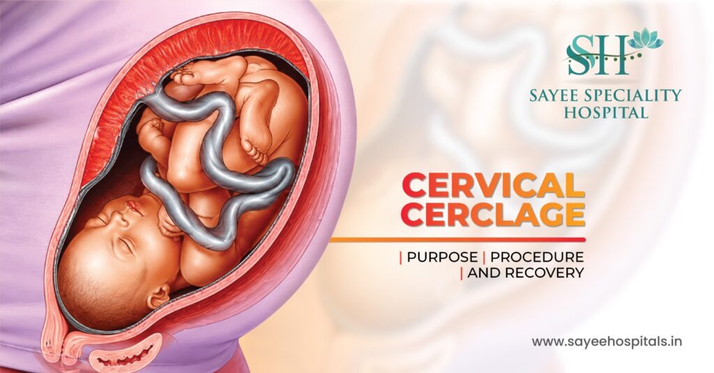
Cervical Cerclage: Purpose,Procedure & Recovery Most people go through pregnancy without ever hearing the term cervical cerclage. For those who do encounter it, it usually arrives at a moment of heightened anxiety, a scan that shows concerning cervical shortening, a history of second-trimester loss that nobody fully explained at the time, or a conversation with a specialist that suddenly reframes the entire pregnancy as higher risk than expected. Understanding what a cerclage actually is, why it’s recommended, and what the procedure involves can take a significant amount of fear out of that experience. It’s a well-established intervention with a clear purpose and a straightforward recovery, and for the pregnancies where it’s indicated, it makes a genuine difference. What the Cervix Does and What Can Go Wrong The cervix is the lower portion of the uterus, a cylindrical structure, typically firm and closed during pregnancy, that keeps the uterine contents secure until the body is ready for labour. In a healthy pregnancy, the cervix remains long, closed, and firm throughout the second and into the third trimester, only beginning to soften, shorten, and dilate as labour approaches. In some pregnancies, the cervix begins to weaken and shorten prematurely, a condition referred to clinically as cervical insufficiency or an incompetent cervix. The cervical tissue loses its structural integrity earlier than it should, and without intervention, this can lead to the cervix opening silently and painlessly during the second trimester, resulting in pregnancy loss or extremely premature delivery. Several factors can contribute to cervical insufficiency. Previous cervical procedures LEEP (loop electrosurgical excision procedure) or cone biopsy for abnormal cervical cells, can reduce the amount of cervical tissue available and affect structural integrity. Previous significant cervical dilation during surgical procedures, uterine abnormalities, connective tissue disorders, and a history of second-trimester losses all raise the index of suspicion. In some cases, cervical shortening is identified incidentally on a routine transvaginal ultrasound during the second trimester, before any symptoms have appeared. Who Needs a Cerclage Cerclage isn’t recommended for every high-risk pregnancy, it’s specifically indicated for cervical insufficiency, and the decision is based on a combination of obstetric history and ultrasound findings. A history-indicated cerclage is placed based on previous pregnancy losses or preterm births consistent with cervical insufficiency, typically planned and placed between weeks 12 and 14 before any cervical change is visible. An ultrasound-indicated cerclage is placed when transvaginal ultrasound during the current pregnancy shows cervical shortening below a certain threshold typically a cervical length below 25mm in a woman with a prior preterm birth. An emergency or rescue cerclage is placed when the cervix is already dilating in the second trimester, which is a more urgent situation with different risks and outcomes than planned placement. The decision involves careful risk-benefit assessment cerclage isn’t without risk, and it’s not beneficial in all situations where preterm birth is a concern. Preterm labour driven by infection or contractions, for instance, is not improved by cerclage and may be worsened by it. The specialist’s judgement about the specific mechanism driving the risk matters enormously. What the Procedure Actually Involves For most women, the anticipation of the procedure is considerably more stressful than the procedure itself. Cervical cerclage is typically performed as an outpatient procedure, meaning the same day return home, usually under spinal or regional anaesthesia rather than general anaesthesia. In the most common approach, the transvaginal McDonald cerclage, the patient is positioned for a standard pelvic examination. The cervix is visualised through a speculum, and strong non-absorbable sutures are placed circumferentially around the cervix, essentially drawing it closed. The procedure takes around 20 to 30 minutes in most cases. A second approach, the Shirodkar cerclage, involves a slightly more involved dissection to place the suture higher on the cervix and is used in certain clinical situations where the McDonald technique isn’t adequate. In rare cases typically where previous transvaginal cerclages have failed, or where the cervix is too short to access vaginally, an abdominal cerclage is performed, either open or laparoscopically, placing the suture at the cervicoisthmic junction above the vaginal portion. This is a more significant procedure and is typically placed before pregnancy or in very early pregnancy. Before the procedure, a transvaginal ultrasound confirms cervical status and foetal wellbeing. Antibiotics are commonly administered to reduce infection risk. Women are usually advised to avoid intercourse for a week before a planned cerclage. The Days After What to Expect Mild cramping and light spotting in the days immediately following the procedure are expected and don’t indicate a problem. Paracetamol is usually sufficient for discomfort management during this period. Most women are monitored for a few hours after the procedure before going home. The recovery window of roughly 7 to 10 days requires some specific restrictions, avoiding strenuous physical activity, heavy lifting, and sexual intercourse while the cervix settles. Beyond that initial period, most women with a cerclage in place return to relatively normal daily activity, though specific guidance varies depending on the clinical situation and the individual obstetrician’s protocol. Regular cervical length monitoring by transvaginal ultrasound continues throughout the pregnancy, typically every two to four weeks, to ensure the cerclage is holding and the cervix isn’t shortening further. Any symptoms of preterm labour, regular contractions, pelvic pressure, increased vaginal discharge, or fluid leakage, warrant immediate assessment rather than watchful waiting at home. The Risks Honest and in Proportion Cerclage is a well-established and generally safe procedure, but it isn’t without risk, and those risks are worth understanding clearly. The most significant potential complication is premature rupture of membranes, where the suture provokes the membranes to break before the pregnancy reaches a viable or term gestation. Infection of the cervix or uterine cavity is another potential complication, which is why antibiotic prophylaxis and monitoring for signs of infection are standard parts of cerclage management. Cervical laceration, tearing at the suture site can occur, particularly if labour begins before the suture is removed. These complications are uncommon, and the risk-benefit calculation for women with a genuine indication
PCOS Is Not Just a Period Problem – Here’s What It’s Actually Doing to Your Body

PCOS Is Not Just a Period Problem – Here’s What It’s Actually Doing to Your Body Most women who get a PCOS diagnosis hear a version of the same explanation. Irregular periods. Cysts on the ovaries. Hormones that are slightly out of balance. Maybe some trouble with weight. Come back if you’re trying to conceive. And then they leave the appointment with a condition that affects roughly one in five women in India – and a picture that is considerably incomplete. PCOS is not primarily a reproductive condition that happens to have some hormonal side effects. It is a complex metabolic and endocrine disorder that uses reproductive symptoms as its most visible calling card. The irregular periods are real and they matter. But they are the part of the iceberg that sits above the water. What sits below it – the insulin resistance, the systemic inflammation, the cardiovascular implications, the mental health burden – is what determines a woman’s long-term health trajectory in ways that have nothing to do with whether she ever wants to get pregnant. That’s the part of the conversation that often doesn’t happen. And it’s the part this article is about. What PCOS Is Actually Doing at the Hormonal Level The name – polycystic ovary syndrome – is somewhat misleading. The ovarian cysts it refers to are not true cysts in the clinical sense. They are small, immature follicles that have failed to complete their development and release an egg, because the hormonal signals that should trigger ovulation are disrupted. The ovaries don’t cause PCOS. They are one of the places where its effects show up. At the core of PCOS is a disruption in the relationship between the pituitary gland, the ovaries, and insulin. The pituitary releases luteinising hormone (LH) in excess, which drives the ovaries to produce more androgens – testosterone and related hormones – than they should. These androgens interfere with the normal follicle development and ovulation cycle. Simultaneously, in around 70 to 80 percent of women with PCOS, the body’s cells are resistant to insulin – meaning the pancreas has to produce more insulin to do the same job, and those elevated insulin levels further stimulate androgen production. The result is a self-reinforcing hormonal loop. Elevated insulin drives more androgens. More androgens disrupt ovulation. Disrupted ovulation means no progesterone is produced in the second half of the cycle, which means oestrogen goes unopposed, and so the hormonal imbalance compounds over time unless something actively intervenes. The Metabolic Consequences Nobody Explains Clearly Type 2 Diabetes Risk Women with PCOS have a significantly elevated lifetime risk of developing type 2 diabetes compared to women without it. Studies consistently put this at three to seven times higher, depending on other risk factors. The pathway is direct: chronic insulin resistance – the foundational metabolic feature of most PCOS cases – is the same mechanism that leads to type 2 diabetes when the pancreas eventually can’t sustain the compensatory insulin output. This isn’t a distant theoretical risk. Women with PCOS who are overweight and have significant insulin resistance can develop impaired glucose tolerance in their thirties. Screening for prediabetes – with a fasting glucose and HbA1c at minimum – should be part of routine PCOS management, not something that only happens when symptoms of diabetes appear. Cardiovascular Disease The cardiovascular implications of PCOS are probably the least discussed and arguably the most consequential. Women with PCOS have higher rates of hypertension, dyslipidaemia (elevated triglycerides and low HDL cholesterol), and systemic inflammation – all of which are independent cardiovascular risk factors. The same insulin resistance that drives the metabolic picture also promotes central adiposity – fat distribution around the abdomen – which is specifically associated with cardiovascular risk over and above general body weight. A woman with PCOS who maintains a relatively normal BMI can still carry a disproportionate amount of visceral fat, which doesn’t show up on a scale but absolutely shows up in cardiovascular risk assessments. Research indicates that women with PCOS have a significantly higher risk of cardiovascular events in midlife. This doesn’t mean it’s inevitable – it means it’s something that warrants active monitoring and preventive management from the time of diagnosis, not when it becomes relevant in the fifties. Endometrial Health Women with PCOS who have infrequent or absent periods are not simply skipping a monthly inconvenience. When ovulation doesn’t occur regularly, progesterone is not produced in the second half of the cycle. Without progesterone to balance it, oestrogen continues to stimulate the endometrium – the uterine lining – month after month without the regular shedding that a period provides. Over time, this unopposed oestrogen stimulation causes the endometrial lining to thicken abnormally, a condition called endometrial hyperplasia. Certain types of hyperplasia carry an elevated risk of progressing to endometrial cancer. This is not a common outcome, but it is a real one – and it’s preventable. Women with PCOS who have fewer than four periods a year should be having regular gynaecological review specifically for this reason. The Visible Effects That Get Treated Separately When They Shouldn’t Be Several PCOS symptoms are routinely managed in isolation – by dermatologists, trichologists, or general practitioners – without the underlying hormonal condition being addressed: Acne Along the Jawline and Lower Face Adult acne in women, particularly when it clusters along the jaw, chin, and lower cheeks, is one of the most consistent dermatological markers of androgen excess. Many women with PCOS spend years cycling through topical treatments and antibiotics for acne that keeps coming back, because the skin is being treated while the hormonal driver continues unchecked. A dermatologist who identifies this pattern and refers for hormonal evaluation is doing exactly the right thing. One who doesn’t is treating a symptom indefinitely. Hair Thinning on the Scalp Androgenic alopecia – hair thinning driven by androgen sensitivity in the hair follicles – presents in women with PCOS as diffuse thinning at the crown and temples rather than the receding hairline pattern seen
Cataract at 35? Why Young People Are Now Walking Into Eye Hospitals
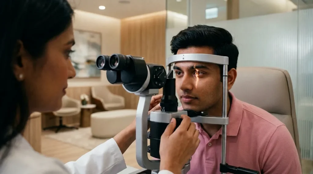
Cataract at 35? Why Young People Are Now Walking Into Eye Hospitals Cataract carries a specific image in most people’s minds. An elderly person, squinting in bright light, moving carefully through a room because the world has gone soft and foggy around the edges. It is, by almost universal assumption, a condition that belongs to the last chapter of life. Which is why it catches people off guard when they’re 34, or 38, or 41 – and an ophthalmologist tells them their lens is clouding. Not severely, not urgently, but enough to explain why night driving has become stressful, why bright lights now come with halos, why reading in dim light has quietly stopped being something they do. Early-onset cataract – cataract developing before the age of 50 – is not a new phenomenon. But it is more common than it used to be, and the reasons why track closely with changes in how younger adults in India live, what conditions they carry, and what their eyes are exposed to across decades of daily life. Understanding this is useful not just for people who already have symptoms, but for anyone in their thirties or forties who has never considered their lens health as something worth thinking about. Because by the time cataract is obvious, it has typically been developing quietly for years. What a Cataract Actually Is The lens of the eye sits just behind the pupil. Its job is to focus light precisely onto the retina at the back of the eye, adjusting its shape for different distances the way a camera lens does. For this to work, the lens needs to be transparent – completely clear, allowing light to pass through without scattering. A cataract is what happens when proteins within the lens begin to clump together and lose their ordered arrangement. Instead of a clear medium, the lens becomes progressively cloudier – like looking through a fogged-up window that can’t be wiped clean from the outside. Light that enters the eye scatters rather than focusing cleanly, producing blurred or hazy vision, increased sensitivity to glare, and in some cases, a yellowing or browning of the visual field. The process is gradual in most cases. A developing cataract in a young person might sit at a level where it’s detectable on examination but doesn’t significantly affect daily function for years. That window – between the cataract forming and it becoming functionally limiting – is exactly when understanding the cause and monitoring progression is most valuable. Why Younger Adults Are Being Diagnosed Earlier Age-related cataract develops because of the cumulative effects of oxidative stress on the lens proteins over decades. In older adults, this is simply a long process reaching its natural conclusion. In younger adults, something is accelerating that process – or initiating a different type of lens clouding altogether. The reasons fall into several distinct categories: Uncontrolled or Long-Standing Diabetes This is the most common medical cause of early cataract in India by a significant margin. High blood glucose over extended periods alters the biochemistry of the lens – specifically, glucose accumulates inside the lens and is converted to sorbitol, which draws water in and disrupts the lens proteins. Diabetic cataracts can develop in patients in their thirties and forties, particularly those whose blood sugar has been poorly controlled for several years. India has one of the highest burdens of diabetes in the world, and a significant proportion of that burden involves people who have had the disease for years before diagnosis – meaning their lenses have been under glycaemic stress without their knowledge. For any diabetic patient, regular eye examinations are not optional. They are part of managing the disease. Long-Term Steroid Use Corticosteroids – used for conditions ranging from asthma and skin disorders to autoimmune diseases and joint problems – are well-established cataract risk factors when used systemically over extended periods. The association is particularly strong with oral steroids, but prolonged use of high-potency topical steroids and nasal steroid sprays also carries a risk, particularly in genetically susceptible individuals. The lens changes induced by steroids tend to occur in the posterior subcapsular region – a specific location that disproportionately affects reading vision and causes significant glare even when the cataract is not yet dense. People using steroids long-term for chronic conditions often don’t connect their worsening reading vision or glare sensitivity to their medication. UV Radiation Exposure Without Protection Ultraviolet radiation – particularly UV-B – induces oxidative damage to the lens proteins. Cumulative, unprotected sun exposure over years is a measurable risk factor for cataract development. This is particularly relevant in a country like India, where outdoor UV intensity is high across most of the year and sunglasses with genuine UV protection are inconsistently used. Sunglasses that are purely cosmetic – tinted but without UV-400 certification – can actually be worse than no sunglasses at all, because the tint causes the pupil to dilate, allowing more UV-laden light to reach the lens. Genuine UV protection requires lenses rated UV-400, which block wavelengths up to 400 nanometres. Trauma to the Eye A direct blow to the eye, a penetrating injury, or even a significant contusion can disrupt the lens capsule and initiate cataract formation. Traumatic cataracts can develop rapidly – within days or weeks of injury – or slowly over years, depending on the nature and severity of the trauma. Sports injuries, accidents, and industrial eye injuries are the most common causes in this category, and many people who sustained eye injuries in their teens or twenties don’t connect them to a cataract appearing a decade later. Other Medical Conditions and Metabolic Factors Hypothyroidism, hyperparathyroidism, atopic dermatitis, and certain genetic conditions are all associated with early lens changes. Nutritional deficiencies – particularly of antioxidant vitamins C and E – may accelerate oxidative lens damage. Heavy, chronic alcohol consumption and smoking are also independent risk factors, with smoking in particular associated with a two-to-threefold increased cataract risk across multiple large studies. Genetic Predisposition A