Thyroid Disorders And Irregular Periods

Thyroid Disorders And Irregular Periods Irregular periods get blamed on a lot of things stress, diet, over-exercising, coming off contraception. And sometimes those explanations are right. But there’s one underlying cause that gets missed surprisingly often, partly because it doesn’t announce itself obviously and partly because the connection isn’t widely known outside clinical settings. The thyroid gland a small butterfly shaped structure sitting at the base of the neck has a reach in the body that extends well beyond what most people associate with it. When it stops working properly, the ripple effects touch almost every system, including the one responsible for menstrual health. For a significant number of women with unexplained irregular periods, the answer isn’t gynaecological at all. It’s sitting in their thyroid function tests. What the Thyroid Does and Why It Matters for Periods The thyroid produces two primary hormones thyroxine (T4) and triiodothyronine (T3) that regulate the body’s metabolic rate. Essentially, these hormones set the pace for how every cell in the body uses energy. Heart rate, body temperature, digestive speed, cognitive function, mood all of these are downstream of thyroid hormone activity. But thyroid hormones don’t operate in isolation. They interact directly with the hormonal architecture of the reproductive system oestrogen, progesterone, and the pituitary hormones that regulate the menstrual cycle, FSH and LH. When thyroid hormone levels drift out of their normal range, that interaction becomes disruptive. The hypothalamic-pituitary-ovarian axis, the hormonal feedback loop that governs ovulation and menstruation is sensitive to thyroid status in ways that produce measurable, sometimes significant changes to the menstrual cycle. There are two directions thyroid dysfunction can go, and they produce opposite effects on periods. Hypothyroidism When the Thyroid Is Underactive Hypothyroidism occurs when the thyroid produces insufficient hormone. The most common cause is Hashimoto’s thyroiditis, an autoimmune condition where the immune system gradually damages thyroid tissue. It can also follow thyroid surgery, radioiodine treatment, or certain medications, and in some parts of the world iodine deficiency remains a significant cause. The systemic effects of hypothyroidism fatigue, weight gain, cold intolerance, constipation, dry skin, hair thinning, brain fog are well known. The menstrual effects are less widely understood but are a consistent feature of the condition. Women with hypothyroidism frequently experience heavy menstrual bleeding menorrhagia. Periods that soak through protection quickly, pass large clots, or last significantly longer than a week are common presentations. The mechanism involves the effect of low thyroid hormone on clotting factors and on the feedback signals that regulate the endometrial lining. Prolonged periods and increased frequency are also seen, the cycle shortening in some women so that periods come every two to three weeks rather than the usual interval. In more severe or longstanding hypothyroidism, periods can become infrequent or stop altogether, as the disruption to the hypothalamic pituitary axis becomes significant enough to suppress ovulation entirely. The broader symptom picture is useful for joining the dots. A woman presenting with heavy, prolonged periods who is also exhausted all the time, gaining weight without dietary changes, struggling with cold, and noticing her hair is thinning deserves thyroid function testing early in the diagnostic workup not as an afterthought once gynaecological causes have been excluded. Hyperthyroidism When the Thyroid Is Overactive Hyperthyroidism sits at the opposite end too much thyroid hormone, driving the body’s systems too fast. Graves’ disease, an autoimmune condition producing stimulating antibodies that activate the thyroid continuously, is the most common cause. Toxic nodular goitre and thyroiditis in its early inflammatory phase are other causes. The systemic picture is the reverse of hypothyroidism weight loss despite normal or increased appetite, heat intolerance, palpitations, anxiety, tremor, excessive sweating, and difficulty sleeping. The menstrual effects are also reversed. Hyperthyroidism tends to suppress menstrual flow rather than amplify it. Light periods, short cycles, infrequent periods, and amenorrhea complete absence of periods are the typical presentations. The high metabolic rate and the effects of excess thyroid hormone on oestrogen metabolism both contribute to a picture where the endometrial lining develops poorly and ovulation becomes irregular or absent. The cardiovascular symptoms of hyperthyroidism the racing heart, the palpitations, the anxiety often dominate the clinical picture and draw attention away from the menstrual changes. But for a woman of reproductive age presenting with light or absent periods alongside those systemic symptoms, hyperthyroidism should be high on the diagnostic list. The Fertility Dimension Thyroid dysfunction affects fertility through several overlapping mechanisms, all of which come back to the same central problem disrupted ovulation. Regular ovulation depends on the precise, coordinated hormonal signalling of the hypothalamic-pituitary-ovarian axis. Thyroid hormone imbalance interferes with that signalling at multiple points. In hypothyroidism, elevated TSH stimulates prolactin secretion, and elevated prolactin suppresses ovulation. In hyperthyroidism, altered sex hormone binding globulin levels change how oestrogen is metabolised and available, disrupting follicular development. The result is that both conditions reduce the frequency of regular ovulation and without consistent ovulation, conception becomes difficult. Studies examining women presenting to fertility clinics consistently find a higher prevalence of thyroid dysfunction than in the general population, and treating the thyroid condition frequently improves both ovulation regularity and conception rates. Beyond conception, untreated hypothyroidism during early pregnancy carries risks of miscarriage, preterm birth, and neurodevelopmental effects on the baby which is why thyroid screening is recommended for women with a history of thyroid dysfunction before and during pregnancy. Women with known Hashimoto’s thyroiditis who are planning to conceive should have their thyroid function optimised before trying. Other Conditions That Overlap One complication of the thyroid-menstruation connection is that several other common conditions produce similar menstrual symptoms, and they can coexist. Polycystic ovarian syndrome (PCOS) causes irregular, infrequent, or absent periods through a completely different mechanism insulin resistance and androgen excess rather than thyroid dysfunction. Endometriosis and uterine fibroids produce heavy, painful periods. Hyperprolactinaemia from causes other than hypothyroidism suppresses ovulation and periods. The practical implication is that a thyroid function test is an essential part of the workup for menstrual irregularity, but a normal thyroid result doesn’t end the
Comprehending Complex Regional Pain Syndrome (CRPS)
Comprehending Complex Regional Pain Syndrome (CRPS) Most pain makes a kind of logical sense. You injure something, it hurts, it heals, the pain fades. The injury and the pain exist in rough proportion to each other, and recovery follows a trajectory that feels predictable even when it’s slow. CRPS doesn’t work like that. It’s one of those conditions that genuinely challenges how most people understand pain, because the pain it produces is entirely disproportionate to whatever caused it, it doesn’t follow a healing curve, and it can persist and intensify long after the original injury has resolved. For the people living with it, that disconnect between what should hurt and what does hurt is one of the most isolating aspects of the condition. What’s Actually Going Wrong Under normal circumstances, the nervous system’s pain response is proportionate and self-limiting. Something damages tissue, the nervous system signals the brain, protective pain behaviour follows, the tissue heals, and the signal quietens. In CRPS, that shutoff mechanism fails. The nervous system either the peripheral nerves, the spinal cord, or the brain’s own processing centres, or some combination of all three becomes dysregulated in a way that sustains and amplifies pain signals far beyond what the original injury warrants. The current understanding describes CRPS as a disorder of neuroinflammation and central sensitisation. The nervous system essentially becomes hypersensitised calibrated to interpret ordinary sensory input as painful, and to produce pain responses that are wildly out of proportion to the actual stimulus. Touch that should feel neutral becomes agonising. A light breeze across the skin can be unbearable. The affected limb develops a life of its own in terms of temperature, colour, and autonomic behaviour changes driven by dysregulation of the sympathetic nervous system running alongside the pain pathways. CRPS is divided into two subtypes based on whether a specific nerve injury is identifiable. Type I previously called reflex sympathetic dystrophy occurs after an injury or illness that didn’t directly damage a named nerve. A wrist fracture, a sprain, a period of immobilisation, these are typical precipitating events. Type II, previously called causalgia, follows a confirmed nerve injury where the damage to a specific nerve is documented. The clinical presentation overlaps significantly between the two, and the distinction matters more for classification than for day-to-day management. It predominantly affects women, with onset most commonly in the fourth decade of life, though it can and does occur across a wide age range including in children and adolescents. What It Feels Like and the Symptoms Beyond the Pain The pain itself is described in terms that communicate how unlike ordinary pain it is. Burning is the most consistent descriptor deep, relentless, often described as feeling like the limb is on fire from the inside. Tearing, stinging, electric these appear frequently in patient descriptions. The intensity is severe enough that CRPS consistently scores among the highest on standardised pain scales, higher than many post-surgical pain states and comparable to active labour. Allodynia is one of the hallmark features pain triggered by stimuli that should cause no pain at all. Light touch, clothing against the skin, a change in air temperature, water from a shower. The threshold for pain drops so dramatically that the affected limb becomes almost impossible to tolerate normal contact with. Beyond the pain, the changes CRPS produces in the affected limb are striking. Skin colour shifts cycling between red, purple, and pale white, sometimes within the same day. Skin temperature changes, with the affected area often markedly warmer or cooler than the surrounding tissue. Skin texture can become thin, shiny, or mottled. Hair and nail growth on the affected limb may accelerate dramatically or slow to almost nothing. Swelling comes and goes. The limb can feel stiff, weak, and impossible to move normally partly because movement hurts, partly because the motor system becomes caught up in the same neurological dysfunction driving the sensory symptoms. All of these features the autonomic skin changes, the allodynia, the disproportionate pain are the nervous system’s dysregulation made visible. They’re not psychological. They’re not imagined. They’re the physiological consequences of a nervous system that’s lost its normal regulatory balance. What Triggers It In the majority of cases, CRPS follows a physical trauma, a fracture is the most common precipitant, with wrist fractures particularly over-represented. Surgery, sprains, crush injuries, burns, and even prolonged immobilisation of a limb have all been documented as triggers. The injury itself is often relatively minor and that’s part of what makes CRPS so disorienting. A fracture that would be expected to heal uneventfully in six to eight weeks instead becomes the starting point of a pain condition that can last years. Why some people develop CRPS following an injury while others with identical injuries don’t remains incompletely understood. Genetic predisposition, pre-existing differences in nervous system sensitivity, inflammatory response characteristics, and psychological factors have all been investigated as contributors. No single predictive factor has been identified. Getting a Diagnosis There’s no blood test, no imaging finding, no single investigation that confirms CRPS. Diagnosis is clinical reached through a combination of the history, the pattern of symptoms, and physical examination findings assessed against the Budapest Criteria, the internationally accepted diagnostic framework for CRPS. The Budapest Criteria require the presence of continuing pain disproportionate to the inciting event, alongside specific combinations of symptoms and signs across sensory, vasomotor, sudomotor, and motor categories. Meeting those criteria in the right clinical context, with other causes excluded, is how the diagnosis is established. Early diagnosis matters significantly. The longer CRPS goes unrecognised and unmanaged, the more entrenched the central sensitisation becomes and the harder it is to treat. A condition that might respond reasonably well to early aggressive rehabilitation becomes considerably more resistant after months or years of untreated neurological dysregulation. Treatment Rehabilitation First, Everything Else in Support Physical and occupational therapy are the foundation of CRPS management, and that priority reflects the underlying neuroscience. The goal isn’t just to manage symptoms, it’s to retrain the nervous system. Given that CRPS
Osgood Schlatter Disease: Managing Pediatric Knee Pain

Osgood Schlatter Disease: Managing Pediatric Knee Pain There’s a particular kind of frustration that comes with being a sporty twelve year old whose knee has decided it’s no longer cooperating. Practice hurts. Games hurt. Kneeling down to tie a shoelace hurts. And the adults around you keep saying “growing pains” in a tone that suggests you should just get on with it. Sometimes, though, those growing pains have a specific name and a specific explanation, a specific management approach, and a genuinely good outcome at the end of it. Osgood-Schlatter disease is one of the most common causes of knee pain in active adolescents, and understanding what’s actually happening makes it considerably less alarming for both the young person experiencing it and the parents watching them limp off the pitch. What’s Actually Going On in the Knee To understand Osgood Schlatter, you need to understand one specific piece of anatomy, the tibial tubercle. It’s the bony bump you can feel just below the kneecap at the top of the shin. This is where the patellar tendon attaches, the tendon that runs from the kneecap down to the shinbone and transmits the force of the quadriceps muscle every time the leg straightens or the knee extends. In adults, that attachment site is solid, mature bone. In growing adolescents, it sits directly over a growth plate, a zone of actively developing cartilage that hasn’t yet hardened into bone. Growth plates are structurally weaker than mature bone, which is what makes them vulnerable. During running, jumping, kicking, and pivoting, essentially the entire vocabulary of most youth sports, the quadriceps muscle contracts powerfully and repeatedly. That contraction pulls on the patellar tendon, which pulls on the tibial tubercle, which pulls on the growth plate underneath it. Do that thousands of times across a training season, and the growth plate gets irritated. The body responds with inflammation swelling, tenderness, and pain concentrated right at that tibial tubercle attachment point. Over time, in some cases, the repeated pulling causes small fragments of bone to form at the site, creating a visible bony prominence that can persist even after the condition resolves. That whole process is Osgood-Schlatter disease. A repetitive stress injury at a structurally vulnerable point in a growing skeleton. Who Gets It and When The peak window is roughly 11 to 14 years old, the years when growth spurts are most rapid and the mismatch between skeletal development and physical demand is most pronounced. During a growth spurt, bones lengthen faster than the surrounding soft tissues, tendons and muscles, can keep pace with. That relative tightness increases the tensile load on the patellar tendon attachment, which is exactly the wrong time to be training hard for a sport that involves a lot of jumping. Boys have historically been more frequently affected than girls, though the gap has narrowed considerably as girls’ participation in high impact sports has increased. Basketball, football, volleyball, gymnastics, and athletics, anything involving repetitive explosive knee extension ,carry the highest association. Both knees are affected simultaneously in roughly a quarter of cases, which can be particularly disruptive to activity. The dominant leg is typically more symptomatic even when both sides are involved. Recognising the Symptoms The pain of Osgood Schlatter is fairly localised and consistent in character, which makes it one of the more recognisable adolescent knee conditions once you know what you’re looking for. The primary symptom is pain and tenderness directly at the tibial tubercle, that bump just below the kneecap. It’s typically sharp during activity and a duller ache at rest. Pressing directly on the bump reproduces the pain reliably, which is part of how clinicians confirm the diagnosis on examination. Kneeling is often particularly uncomfortable because it places direct pressure on the affected area. The pattern follows activity closely. Symptoms intensify during and immediately after sports running, jumping, squatting and ease with rest. That activity rest relationship is a consistent feature and helps distinguish Osgood-Schlatter from other causes of adolescent knee pain. In some children, the tibial tubercle becomes visibly enlarged, a bony protrusion that’s noticeable even when the leg is extended. This is the fragment of bone that forms in response to repeated pulling at the growth plate, and once it forms it tends to persist even after the pain has fully resolved. Tightness in the quadriceps and hamstrings is commonly found alongside the pain partly as a consequence of the body’s protective guarding response around a painful joint, and partly because tight muscles increase the tensile load on the tendon attachment and contribute to the problem. Getting the Diagnosis In most cases, the diagnosis is clinical the combination of the child’s age, the sport they play, the location and character of the pain, and the tenderness on palpation of the tibial tubercle is enough. No imaging required. X-rays are occasionally ordered, primarily to rule out other causes of adolescent knee pain, like a stress fracture of the tibial tubercle or a bone tumour, both of which are rare but worth excluding when the presentation is atypical. X-ray can also visualise calcification or fragmentation at the tubercle in established cases, though this doesn’t change management. The important differential to keep in mind is that not all anterior knee pain in adolescents is Osgood-Schlatter. Sinding-Larsen-Johansson syndrome, a similar apophysitis at the lower pole of the patella rather than the tibial tubercle, produces pain slightly higher up and is occasionally confused with it. Patellofemoral pain syndrome presents differently but overlaps in age group and activity association. A clinician with experience in paediatric musculoskeletal conditions will work through these distinctions. Managing It the Approach That Actually Works Treatment is conservative in almost every case, and the core principle is straightforward: reduce the load on the irritated growth plate until it matures and the vulnerability resolves. Relative rest is the starting point not complete cessation of all activity, which is both impractical and unnecessary, but a reduction in the high-impact activities driving the symptoms. Most clinicians recommend modifying rather than
Exanthem : Viral Rashes

Exanthem : Viral Rashes Most parents have been there, you’re managing what seems like a straightforward fever, and then on day two or three, a rash appears. The instinct is immediate: panic, Google, more panic. Is it serious? Is it contagious? Does it change everything about how this illness needs to be handled? Sometimes the rash is the most useful piece of information in the whole picture. In viral illness, a skin eruption isn’t just an inconvenient side effect, it’s often the body communicating exactly what’s going on underneath. What a Viral Exanthem Actually Is The word exanthem comes from the Greek for “breaking out” and that’s essentially what it describes. A viral exanthem is a widespread skin eruption that develops as a secondary response to a viral infection. It’s not the infection itself appearing on the skin, it’s the consequence of the body’s interaction with the virus, and it can look quite different depending on which virus is responsible. The mechanism varies. In some cases, the rash is the immune system’s work inflammatory cells and immune mediators reaching the skin as part of the broader defence response. In others, the virus directly infects skin cells, causing localised damage that becomes visible. In others still, viral toxins trigger a skin reaction. The same visible outcome spots, blotches, bumps can arise through different underlying pathways, which is partly why identifying the specific virus from the rash alone isn’t always straightforward. Viral exanthems are most common in children, largely because adults have typically already built immunity to the viruses most associated with them through prior exposure or vaccination. The classic childhood illnesses chickenpox, measles, roseola, and fifth disease, all produce characteristic rash patterns that have been documented for long enough that experienced clinicians can often identify them on appearance alone. More recently, COVID-19 joined the list of viruses known to produce cutaneous eruptions, in patterns ranging from urticarial rashes to chilblain-like changes on the extremities. What It Looks Like and How It Spreads Across the Body One of the clinically useful patterns with viral exanthems is the direction of spread. Most begin centrally on the face or the trunk, before moving outward to the limbs. That centrifugal progression is a consistent feature across many viral rashes and helps distinguish them from other causes of skin eruption. The character of the rash varies considerably by cause. Chickenpox produces intensely itchy, fluid-filled vesicles that appear in crops new lesions arriving while older ones are crusting over, so multiple stages coexist simultaneously. Measles produces a flat, blotchy maculopapular rash that spreads from the hairline downward, preceded by the distinctive Koplik’s spots inside the mouth. Roseola, caused by human herpesvirus 6, follows a particular sequence high fever for several days, then as the fever breaks, a rose pink rash appears across the trunk. Fifth disease, caused by parvovirus B19, gives the characteristic “slapped cheek” appearance before spreading to the limbs in a lacy pattern. Some viral rashes are itchy enough to be genuinely distressing, particularly chickenpox in children. Others cause no discomfort at all. The sensation or lack of it doesn’t indicate severity. The Systemic Symptoms That Come With It The rash rarely arrives in isolation. Because the underlying cause is a systemic viral infection, the skin changes sit alongside the broader picture of the illness and those accompanying symptoms are often more informative than the rash itself. Fever is almost universal, and in some viral exanthems it runs high enough to be concerning in young children from a febrile convulsion risk standpoint. Headache, general malaise, and the bone-tired fatigue that comes with viral illness are consistent companions. Upper respiratory symptoms runny nose, sore throat, conjunctivitis are prominent in measles and several other viral exanthems. Loss of appetite and abdominal discomfort appear frequently enough to occasionally muddy the picture and send parents down the gastroenteritis pathway before the rash appears. The full symptom cluster, taken together with the rash pattern and the child’s age and vaccination history, is usually what points toward the specific diagnosis. Contagion the Part That Matters for Everyone Around the Child This is where the distinction between the rash and the virus matters enormously. The rash itself isn’t contagious through touch pressing on a chickenpox blister, for instance, doesn’t transmit the varicella virus to an intact skin surface. But the virus causing the rash absolutely is contagious, typically through respiratory droplets or, in the case of chickenpox, through direct contact with blister fluid. Contagion windows vary by virus and don’t always align neatly with the rash. Measles, for instance, is contagious from around four days before the rash appears to four days after meaning a child is spreading the virus during the prodromal phase when nobody yet knows measles is what they’re dealing with. Fifth disease is actually most contagious before the rash develops, which is similarly counterintuitive. The groups requiring particular caution are pregnant women and immunocompromised individuals. Parvovirus B19 fifth disease can cause severe anaemia in a developing fetus if a non-immune woman is exposed during pregnancy. Measles and chickenpox carry significantly elevated complication risks in immunocompromised children and adults. If a child with a viral exanthem has had contact with either of these groups, that’s worth flagging to a healthcare provider promptly rather than assuming it’ll be fine. Managing It at Home For the majority of viral exanthems in otherwise healthy children, treatment is supportive there’s no antiviral medication that targets most of these viruses directly, and the illness needs to run its course. The goal is keeping the child comfortable while the immune system does its job. For itching particularly significant with chickenpox cool compresses applied to affected areas provide temporary relief. Calamine lotion remains a time-tested option for itchy rashes, and mild topical hydrocortisone cream can reduce localised inflammatory itch. Keeping nails short and clean reduces the damage from scratching, which matters because broken skin from scratch wounds creates an entry point for bacterial infection, a secondary complication that can turn a straightforward viral illness into
Postpartum Hemorrhage: Understanding And Managing Excessive Bleeding.
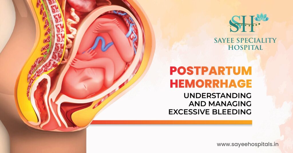
Postpartum Hemorrhage: Understanding And Managing Excessive Bleeding. Childbirth is one of those experiences where the focus understandably stays fixed on the baby. The room shifts, attention moves, and the mother’s ongoing physical state can quietly take a back seat in those first overwhelming hours. Which is part of why postpartum hemorrhage catches so many families off guard. The birth went well. The baby is here. And then something goes wrong anyway. PPH is one of the leading causes of maternal mortality globally, and yet awareness of it outside clinical settings remains surprisingly low. Understanding what it is, why it happens, and what the warning signs look like after leaving hospital isn’t alarmist, it’s genuinely important. What Postpartum Hemorrhage Actually Means Some degree of bleeding after delivery is normal and expected. The body loses blood during every birth, the clinical threshold that defines postpartum hemorrhage is blood loss exceeding one litre after delivery, or any amount of blood loss accompanied by signs that the body is struggling to compensate, such as a falling blood pressure or a rising heart rate. That physiological distinction matters. PPH isn’t defined purely by volume, it’s defined by what that volume does to the patient. A fit, healthy woman with a higher blood volume may tolerate a loss that would push a smaller or more anaemic woman into shock. The vital signs tell the fuller story. PPH is categorised by timing. Primary PPH occurs within the first 24 hours after delivery and the majority of cases, in practice, develop within the first hour after birth, which is why that immediate postpartum period is so closely monitored in clinical settings. Secondary or late PPH occurs anywhere from 24 hours to 12 weeks postpartum. This delayed presentation is the one more likely to catch families off guard at home, when the birth feels long past and significant bleeding is the last thing expected. The Four Ts How Clinicians Think About the Cause When PPH occurs, the clinical priority is identifying the source of bleeding as quickly as possible, because treatment is entirely different depending on what’s causing it. The Four Ts framework — Tone, Trauma, Tissue, Thrombin gives care teams a systematic way to work through the possibilities rapidly. Tone refers to uterine atony, the failure of the uterine muscle to contract adequately after delivery. In a normal third stage of labour, the uterus contracts firmly, compressing the blood vessels where the placenta was attached and limiting blood loss. When those contractions are weak or absent, those vessels remain open. Uterine atony accounts for around 80% of PPH cases, making it by far the most common cause and the first thing clinicians assess. Trauma covers physical injury to the birth canal lacerations of the vagina or cervix, or in more serious cases, uterine rupture. These are particularly likely following instrumental deliveries, prolonged labour, or when a baby is larger than average. Unrepaired tears bleed continuously regardless of how well the uterus is contracting, which is why a thorough examination of the birth canal follows every delivery. Tissue refers to retained products of conception fragments of placenta or membranes that remain attached to the uterine wall rather than delivering completely. The uterus cannot contract properly around retained tissue, and those fragments themselves can be a source of ongoing bleeding. Ultrasound after delivery can identify retained placental material that isn’t clinically obvious. Thrombin covers coagulopathy conditions affecting the blood’s ability to clot. These may be pre-existing clotting disorders that were known before pregnancy, or they may develop acutely during a complicated labour. Disseminated intravascular coagulation, or DIC, is a serious acquired coagulopathy that can develop in the context of severe PPH itself, creating a dangerous cycle where bleeding drives coagulopathy which drives further bleeding. Recognising the Signs Especially After Going Home In the immediate postpartum period, hospital staff are monitoring closely enough that early PPH is generally identified and managed before it escalates. The more vulnerable window for many women is after discharge, when the clinical oversight is gone and the responsibility for recognising warning signs shifts to the patient and her family. Heavy lochia the normal postpartum vaginal discharge, is expected in the days after birth, gradually reducing over the following weeks. The signs that something has shifted beyond normal include soaking through a maternity pad within an hour or less, passing blood clots larger than roughly the size of a golf ball, or noticing that bleeding has increased rather than gradually decreased. Systemic signs of significant blood loss include dizziness, light headedness, or fainting particularly on standing. Blurred vision, confusion, or a feeling of unreality. Skin that looks pale and feels cold or clammy. A heart rate that feels noticeably rapid or irregular. These are the body’s compensatory responses to falling blood volume they’re the signs that something is happening that goes beyond normal postpartum recovery. Any of these, individually or together, warrants calling for emergency help rather than waiting to see if things improve. Secondary PPH presenting at home can escalate quickly, and the response time matters. How It’s Treated Management is driven by cause, and in most cases several interventions happen simultaneously rather than sequentially. For uterine atony the most common cause, the immediate priorities are stimulating uterine contraction and replacing lost blood volume. Uterotonic medications, primarily oxytocin, are given to prompt the uterine muscle to contract. Ergometrine, misoprostol, and tranexamic acid are additional agents used depending on clinical circumstances. Uterine massage bimanual compression performed by the clinical team physically stimulates contraction when medication alone isn’t achieving it. Traumatic bleeding from lacerations or tears is managed surgically suturing the damaged tissue to achieve haemostasis. Retained placental tissue may require manual removal or surgical evacuation under anaesthesia. For severe or rapidly progressing hemorrhage, resuscitation runs alongside haemostasis efforts. IV fluids maintain circulating volume while the team works to control the source. Blood transfusion often multiple units replaces lost red cells and clotting factors. In major hemorrhage, the ratio of packed red cells to fresh frozen plasma
Revolutionizing Recovery Through Multimodal Pain Management
Revolutionizing Recovery Through Multimodal Pain Management For a long time, the default response to post surgical pain was straightforward: strong opioids, high doses, and the assumption that serious surgery meant serious medication. That approach worked in the narrow sense, it controlled pain, but it came with a set of costs that the medical community has spent the last two decades reckoning with. Dependence, prolonged sedation, delayed recovery, constipation, nausea, and a difficult transition off medication once the acute phase was over. Modern surgical recovery looks quite different now. Not because pain is being taken less seriously but because the understanding of how to treat it has become genuinely more sophisticated. Why One Drug Was Never Really the Answer Pain isn’t a single signal travelling along a single pathway. It’s a complex cascade involving multiple sites peripheral nerve endings at the surgical site, the spinal cord where signals are relayed and modulated, and the brain where they’re ultimately interpreted. Targeting only one point in that system with one type of medication was always going to be an incomplete solution. Multimodal analgesia – MMA works on a different principle entirely. Instead of one drug at high dose, it combines several medications with different mechanisms of action at lower individual doses. Paracetamol addresses central pain processing. NSAIDs reduce peripheral inflammation at the surgical site. Gabapentinoids calm sensitised nerve pathways. Local anaesthetics delivered through regional techniques block transmission at the nerve level. Each works on a different part of the pain pathway simultaneously, and the combined effect is greater than any single agent could achieve alone. The practical result is meaningful better pain control with significantly lower opioid requirements. And lower opioid requirements mean fewer opioid side effects, clearer cognition during recovery, faster return of gut function, earlier mobilisation, and reduced risk of the kind of prolonged opioid use that can follow major surgery when high-dose prescribing is the norm. Regional Anaesthesia Targeting the Source One of the most significant shifts in surgical pain management over the last decade has been the expanded use of regional anaesthesia techniques. Rather than relying solely on systemic medication that travels through the bloodstream and affects the whole body, regional techniques deliver anaesthetic directly to the nerve or nerve group supplying the surgical site. Nerve blocks injecting local anaesthetic around a specific nerve can provide hours to days of targeted pain relief from a single procedure. For hip and knee replacements, femoral nerve blocks and adductor canal blocks have become standard parts of the recovery protocol in many centres. For shoulder surgery, interscalene blocks. For abdominal procedures, transversus abdominis plane blocks. The specificity is remarkable patients can be alert, oriented, and relatively comfortable while the operative area remains effectively numb. Epidural analgesia, well established in obstetric care, has an equally important role in major abdominal and thoracic surgery, providing sustained segmental pain control that allows earlier breathing exercises and earlier mobilisation after procedures where systemic opioids would otherwise be heavily sedating. These techniques sit at the centre of Enhanced Recovery After Surgery protocols the structured, evidence-based frameworks that major surgical centres now use to systematically reduce surgical stress, minimise complications, and accelerate the return to normal function. The Team Behind the Plan What makes modern perioperative pain management work isn’t just the pharmacology, it’s the coordination. A well-functioning team brings together distinct expertise at each stage of the surgical journey, and the quality of that coordination directly affects patient outcomes. Surgeons and anaesthesiologists develop the individualised plan before the patient reaches the operating table identifying the most appropriate regional techniques, selecting an opioid-sparing analgesic combination suited to the procedure and the patient’s medical history, and anticipating where the pain management challenges are likely to arise. Nurses are the continuous thread through the recovery process. They’re monitoring pain scores regularly, identifying when the plan needs adjustment, recognising early signs of complications, and doing the patient education work that makes discharge safe rather than just timely. A patient who understands what level of discomfort is expected, what to take and when, and what symptoms should prompt them to seek help is a patient who manages their recovery at home far more successfully. Pharmacists contribute precision reviewing the complete medication picture, identifying interactions, ensuring the doses being prescribed are appropriate for the individual’s weight, renal function, and other medications. In complex patients, that review catches problems that might otherwise only surface as complications. What Happens Before the Surgery Matters Effective pain management doesn’t start in the recovery room. It starts in the pre-operative consultation, and the evidence strongly supports that patients who are properly prepared before surgery recover more smoothly after it. Preoperative education sets realistic expectations which sounds simple but has measurable clinical impact. Patients who know in advance what the normal trajectory of post-surgical discomfort looks like, what their analgesic plan involves, and what non-pharmacological tools are available to them are less anxious going into surgery, report lower pain scores postoperatively, and require less rescue medication. Anxiety and pain are physiologically intertwined in ways that aren’t just psychological the stress response amplifies pain perception, and reducing preoperative anxiety reduces postoperative pain. Non-pharmacological techniques introduced preoperatively become practical tools during recovery. TENS transcutaneous electrical nerve stimulation delivers low-level electrical impulses through the skin that interfere with pain signal transmission and provide genuine, if modest, relief as part of a broader plan. Cryotherapy structured cold application to the surgical site, reduces local inflammation and provides localised pain relief in the early post-operative period. Breathing exercises and relaxation techniques give patients agency over their own comfort during a phase when a lot feels out of their control. Scheduled Dosing a Small Change With a Big Effect One of the most practically important shifts in post-surgical pain management is the move from as needed dosing to scheduled dosing for non-opioid analgesics. It sounds like an administrative detail. It isn’t. When medication is taken only when pain becomes severe, there are repeated cycles of under-treatment followed by catch up, the pain climbs, the
The Road To Recovery: Mastering Non-Surgical Joint Pain Relief
The Road To Recovery: Mastering Non-Surgical Joint Pain Relief Somewhere along the way, a lot of people picked up the idea that chronic joint pain has two settings put up with it, or get surgery. That binary isn’t accurate and for many patients, it leads to years of unnecessary suffering on one end, or jumping to an invasive procedure before less risky options have been properly explored on the other. The reality is that conservative, non surgical management of joint pain has become genuinely sophisticated. For knees, hips, and shoulders dealing with arthritis or chronic degeneration, the toolkit available without going anywhere near an operating theatre is broader and more effective than most people realise. Starting With the Pain Itself Before anything else can happen before rehabilitation, before lifestyle changes, before any of the longer term work, the pain needs to be at a level where movement is actually possible. That’s not a luxury consideration. It’s the clinical prerequisite for everything that follows, because a person in severe pain won’t and can’t do the physiotherapy that drives real recovery. For mild to moderate joint pain, non prescription anti inflammatories like ibuprofen or naproxen are a reasonable starting point. They address the inflammatory component of joint pain rather than just masking the sensation, which makes them more useful than simple analgesics for arthritis type conditions. Paracetamol works differently, it doesn’t reduce inflammation, but remains a useful baseline pain management tool, particularly for people who can’t tolerate NSAIDs due to gastrointestinal or cardiovascular considerations. When oral medication isn’t providing sufficient relief, targeted injections bring the treatment directly to the source. Corticosteroid injections are the most established option anti inflammatory medication delivered directly into the joint space, bypassing the systemic effects of oral steroids. The pain relief they produce can be significant and can last anywhere from several weeks to several months, creating a window where meaningful rehabilitation becomes possible. They’re not a long-term solution on their own, but as a bridge to getting someone moving and strengthening, they earn their place. Visco supplementation uses hyaluronic acid, a substance that naturally occurs in healthy joint fluid injected into the joint to improve lubrication and cushioning. The evidence is more mixed than for corticosteroids, but a meaningful subset of patients, particularly those with knee osteoarthritis, report sustained benefit. It’s a reasonable option to consider when steroid injections haven’t provided adequate duration of relief. PRP therapy platelet rich plasma sits in the regenerative medicine space and has attracted significant interest over the last decade. The process involves drawing a small amount of the patient’s own blood, concentrating the platelets through centrifugation, and injecting that concentration back into the affected joint. Platelets carry growth factors that play a role in tissue repair, and the theory is that a high local concentration accelerates the body’s natural healing response. The evidence base is still developing, but results in knee osteoarthritis and certain tendon related conditions are promising enough that it’s increasingly offered as a serious option rather than an experimental one. Physical Therapy, The Part That Actually Changes the Trajectory If there’s one intervention that consistently makes the most difference to long-term joint pain outcomes, it’s structured physiotherapy. Not generic exercise, not a leaflet of stretches, a properly tailored programme designed around the specific joint, the specific pattern of weakness and restriction, and the specific goals of the individual patient. The logic is straightforward. Joints don’t exist in isolation. The knee is supported by the quadriceps, hamstrings, glutes, and hip stabilisers. The shoulder depends on the rotator cuff, the periscapular muscles, and the deep stabilisers of the glenohumeral joint. When the muscles surrounding a joint are weak or poorly coordinated, the joint itself absorbs disproportionate load, which accelerates degeneration and worsens pain. Rebuilding that muscular support doesn’t just reduce pain. It changes the mechanical environment of the joint in ways that slow down the underlying condition. Range of motion work matters alongside strengthening. Joints that are protected through inactivity avoided because movement hurts lose flexibility progressively, which creates its own layer of stiffness and dysfunction on top of the underlying problem. Gentle, progressive mobility work alongside strengthening is what restores function rather than just managing symptoms. A good physiotherapist also addresses movement patterns, the way a person walks, climbs stairs, or performs daily tasks, that may be loading the joint inefficiently. Small corrections to mechanics can have surprisingly large effects on pain over time. Weight, Activity, and the Things You Can Control Lifestyle modification gets mentioned frequently in this context and sometimes gets dismissed as generic advice. It isn’t. The mechanical reality of what body weight does to weight-bearing joints is significant and well documented. Each kilogram of body weight translates to roughly three to four kilograms of force across the knee joint during walking, and more during stairs or slopes. For someone carrying excess weight with knee or hip osteoarthritis, even a modest reduction in body weight produces a disproportionately large reduction in joint load. That’s not about aesthetics, it’s basic biomechanics, and the clinical evidence supporting weight loss as a joint pain intervention is solid. Activity modification doesn’t mean stopping activity. It means substituting high impact loading for lower-impact alternatives that maintain cardiovascular fitness and muscle mass without the joint stress. Swimming is the most joint friendly option available, the water offloads the joints almost entirely while allowing real muscular work. Cycling, both static and outdoor, provides excellent lower limb strengthening with minimal impact. These aren’t consolation prizes for people who can’t run they’re genuinely effective forms of exercise that many people find they prefer once they try them properly. Assistive devices knee braces, walking aids, custom orthotics, get underused because people associate them with a kind of defeat. That framing doesn’t serve anyone. A well fitted knee brace that offloads the medial compartment reduces pain and allows more activity, which maintains muscle mass, which improves the joint environment. An orthotic that corrects overpronation changes how load is distributed all the way up through the
Understanding Congenital Heart Defects: Protecting Your Child’s Heart Health
Understanding Congenital Heart Defects: Protecting Your Child’s Heart Health Finding out your child has a heart condition is one of those moments that reshapes everything. Whether it comes during a routine anomaly scan, in the hours after birth, or later in childhood when symptoms start to surface the words “congenital heart defect” land heavily, regardless of how they’re delivered. And the first thing most parents want to know, before anything else, is what it actually means for their child. The answer varies enormously depending on the specific defect. Some are minor, self resolving, and require nothing more than monitoring. Others are complex, require surgery, and demand lifelong medical involvement. Understanding the landscape, what these conditions are, how they’re recognised, and what modern cardiac care actually offers, is the starting point for navigating all of it. What a Congenital Heart Defect Actually Is The heart begins forming in the first six weeks of pregnancy remarkably early, before most women even know they’re pregnant. It starts as a simple tube and goes through an elaborate developmental process, folding, dividing, and forming chambers, valves, and vessels. Congenital heart defects occur when some part of that process doesn’t complete correctly. The result is a structural abnormality that’s present from birth, ranging from a small hole between chambers to complex malformations involving multiple structures simultaneously. CHDs are the most common type of birth defect globally, affecting roughly 1 in 100 births. That prevalence makes them far more common than most people realise and the spectrum of severity is wide enough that “congenital heart defect” as a category encompasses everything from conditions that close on their own in infancy to conditions requiring open heart surgery in the first days of life. The Main Types What’s Structurally Different Broadly, congenital heart defects fall into a few structural categories, though individual conditions often involve more than one type of abnormality. Septal defects holes in the walls dividing the heart’s chambers are among the most common. An atrial septal defect (ASD) is a hole between the two upper chambers. A ventricular septal defect (VSD) is a hole between the two lower chambers. Both allow oxygenated and deoxygenated blood to mix in ways that reduce the heart’s efficiency. Small VSDs frequently close on their own as the child grows. Larger defects in either category typically require intervention. Valve abnormalities affect how blood moves between chambers or out into the major vessels. Stenosis, where a valve is too narrow to open fully forces the heart to work harder to push blood through the restricted opening. Valve regurgitation, where a valve fails to close properly and allows blood to leak backward, creates a different kind of inefficiency. Ebstein’s anomaly, which affects the tricuspid valve, is one example of a more complex valve malformation. Complex conditions involve multiple structural abnormalities working together to significantly disrupt normal blood flow. Tetralogy of Fallot, one of the most well-known complex CHDs involves four simultaneous abnormalities, including a large VSD and pulmonary stenosis, that together cause significant oxygen desaturation. Hypoplastic left heart syndrome involves severe underdevelopment of the left side of the heart, requiring a series of staged surgeries in the first years of life. Transposition of the great arteries, where the aorta and pulmonary artery are switched, is another serious condition requiring surgery shortly after birth. Recognising the Signs, In Newborns and Older Children Serious defects are often identified before birth during anomaly ultrasound scans, or detected shortly after delivery during newborn examination. But not all CHDs are obvious immediately, and some are picked up later in childhood. In newborns and young infants, the signs worth knowing include cyanosis, a blue or grey tinge to the lips, skin, or fingernails indicating insufficient oxygen in the bloodstream. Rapid or laboured breathing, particularly at rest, is another significant indicator. Swelling around the eyes, abdomen, or legs suggests the heart isn’t circulating fluid efficiently. Poor feeding and inadequate weight gain where a baby tires quickly during feeds and takes in insufficient calories as a result, can indicate that the heart is working too hard to sustain normal activity. In older children, symptoms shift. Fainting or near fainting during physical activity, tiring far more easily than peers, unexplained breathlessness, and swelling in the hands and feet during exertion are all presentations worth investigating. Some children with undiagnosed CHDs are identified during routine screening or incidentally on imaging done for another reason. What Increases the Risk For the majority of congenital heart defects, no single identifiable cause is found. Most arise from a combination of genetic predisposition and environmental factors during early fetal development. Several maternal health factors during pregnancy are associated with increased CHD risk. Rubella infection in the first trimester carries a well-established link to cardiac defects, which is precisely why rubella vaccination before pregnancy matters. Poorly controlled pre gestational diabetes increases the risk of several cardiac malformations. Certain medications taken during the first trimester — including lithium, isotretinoin, and some anticonvulsants, are associated with cardiac abnormalities and require careful risk-benefit discussion before and during pregnancy. Smoking and alcohol during pregnancy both carry cardiac developmental risks alongside their broader fetal effects. Genetic conditions including Down syndrome (trisomy 21), Turner syndrome, and DiGeorge syndrome all carry significantly elevated rates of congenital heart abnormalities, which is why cardiac assessment is a standard part of the workup when these conditions are identified. From a prevention standpoint, consistent prenatal care, folic acid supplementation from preconception onward, avoiding harmful substances, and ensuring vaccination status is current before pregnancy are all meaningful steps, not guarantees, but genuine risk reduction measures. Treatment From Monitoring to Surgery What treatment looks like depends entirely on the specific defect and its severity. Some small defects particularly small VSDs and small ASDs are monitored rather than treated, with regular echocardiography tracking whether they’re closing spontaneously as the child grows. Many do, particularly in the first few years of life. Catheter-based interventions have transformed the management of several defect types. Rather than open surgery, a thin tube is guided through
Regular Cervical Health Screening Is a Life saving Choice
Regular Cervical Health Screening Is a Life saving Choice There’s a particular kind of medical appointment that women consistently put off not because they don’t know it matters, but because it feels uncomfortable, awkward, or easy to reschedule when nothing seems wrong. Cervical screening sits firmly in that category for a lot of people. And the uncomfortable truth is that “nothing seems wrong” is exactly the problem, because early cervical changes almost never produce symptoms. By the time something feels wrong, the window for easy intervention has often already closed. Cervical screening is one of the few medical tests that doesn’t just detect cancer early, it can prevent it from developing at all. That distinction is worth understanding properly. What Screening Is Actually Looking For The vast majority of cervical cancers somewhere above 99% are caused by persistent infection with high risk strains of Human Papillomavirus, or HPV. HPV is extraordinarily common. Most sexually active people will encounter it at some point, and most of the time the immune system clears it without any intervention or even awareness. The problem arises when a high risk strain persists and, over years, begins causing abnormal changes in the cells lining the cervix. Those changes called dysplasia or cervical intraepithelial neoplasia are precancerous. Left undetected and unmanaged, some will progress to cervical cancer. Detected early, they’re highly treatable. That’s the window screening is designed to exploit. HPV infection to cervical cancer typically takes ten to fifteen years. That’s a long runway long enough for regular screening to catch the process well before it becomes dangerous, in almost every case. The Two Main Tests and What Each One Does There are two primary screening tools, and understanding the difference between them helps make sense of why recommendations are structured the way they are. The Pap smear or Pap test, named after the physician Georgios Papanicolaou who developed it involves collecting a small sample of cells from the surface of the cervix during a speculum examination. Those cells are examined under a microscope for abnormal changes. It’s been the foundation of cervical screening programmes for decades and has dramatically reduced cervical cancer mortality in countries where it’s consistently implemented. The HPV test takes the same sample but analyses it differently specifically looking for the presence of high risk HPV strains rather than examining cell morphology. The logic is that HPV is the cause of almost all cervical cancers, so testing for the virus directly is in many ways a more upstream approach. A negative HPV test provides strong reassurance that significant cell changes are unlikely to be developing. Co testing combines both in a single appointment, providing the most comprehensive picture identifying both existing cell changes and the viral presence that drives them. Many guidelines now support primary HPV testing as the preferred approach, with the Pap smear following as a reflex test if HPV is detected. If either test returns an abnormal result, the next step is typically colposcopy, a procedure where a specialist examines the cervix under magnification using a colposcope, essentially a specialist microscope used externally. If suspicious areas are identified, a small biopsy is taken for laboratory analysis. It sounds intimidating, but colposcopy is an outpatient procedure and provides the definitive information needed to decide whether and what treatment is required. When to Screen and How Often Screening guidelines vary somewhat between countries, but the broad framework is consistent. For women aged 21 to 29, a Pap test every three years is the standard recommendation. HPV testing alone isn’t generally recommended in this age group because HPV infections are common in younger women and typically clear on their own testing for HPV too early generates a lot of findings that would resolve without intervention, causing unnecessary anxiety and follow up. From age 30 to 65, the options widen. HPV testing alone every five years, co-testing every five years, or a Pap smear every three years are all accepted approaches. The five year interval for HPV-based testing reflects the strong reassurance a negative HPV result provides, if high risk HPV isn’t present, the risk of significant cell changes developing within that window is very low. Beyond 65, women who have had consistently normal results and no recent abnormal history can typically stop screening, on the basis that their risk profile no longer justifies the continuation of routine testing. This decision should always be made in conversation with a clinician rather than assumed. Women with certain risk factors, a history of cervical changes, HIV, immunosuppression, or diethylstilbestrol exposure before birth, may need more frequent screening than the standard schedule. Individual circumstances always take precedence over general guidelines. Getting the Test Right, Simple Preparation Steps Pap smear accuracy depends partly on the quality of the cell sample collected, and there are a few straightforward things that reduce the chances of an inconclusive result. For two days before the test, avoid sexual intercourse, vaginal douching, tampons, and any vaginal creams or medications. These don’t make the test impossible, but they can interfere with the cell sample in ways that reduce clarity. Testing during a menstrual period is technically possible but ideally avoided if the timing can be adjusted, a heavy bleed can obscure the sample. The test itself takes a matter of minutes. A speculum is used to visualise the cervix, a small brush or spatula collects cells from the cervical surface, and that’s essentially it. Mild cramping or light spotting afterward is normal and resolves quickly. The discomfort is real for some people particularly those with vaginismus, a history of trauma, or other anatomical considerations and it’s always worth telling your clinician if you have concerns beforehand, as there are ways to make the experience more manageable. HPV Vaccination the Prevention Layer Screening and vaccination work together rather than replacing each other. The HPV vaccine offered routinely to adolescents in most countries’ immunisation, programmes protects against the high risk strains most commonly responsible for cervical cancer, particularly HPV 16 and 18, which together
Conquering Cancer Pain : Strategies For Comfort And Relief

Conquering Cancer Pain : Strategies For Comfort And Relief Pain is one of those aspects of cancer that doesn’t get talked about as openly as it should. People discuss diagnoses, treatment plans, survival statistics, but the day to day reality of living with cancer pain, and what can actually be done about it, often stays in the background. That silence does real harm, because cancer pain is one of the most treatable aspects of the disease and yet it remains significantly undertreated in a large number of patients. That gap between what’s possible and what people are actually experiencing deserves to be closed. Why Cancer Pain Is Different for Everyone Cancer pain isn’t one thing. It’s a broad category that encompasses entirely different types of pain depending on what’s causing it, where it’s located, and what stage the disease is at. Some patients describe a constant dull ache that never fully leaves. Others experience sharp, sudden flares that arrive unpredictably. Burning, pressure, stabbing, throbbing, these are all words that appear in cancer pain descriptions, sometimes from the same patient on different days. The source of the pain matters for how it’s treated. Pain caused directly by the tumour, a mass pressing on a nerve, infiltrating bone tissue, or compressing an organ, behaves differently from treatment-related pain. Chemotherapy-induced peripheral neuropathy, for instance, produces burning or tingling sensations in the hands and feet that result from nerve damage caused by certain chemotherapy agents. Post-surgical pain involves tissue healing. Radiation-related pain can involve inflammation of the treated area. Understanding which mechanism is driving the pain is the first step toward treating it effectively rather than just reaching for the nearest painkiller. The Treatment Toolkit What’s Actually Available Modern cancer pain management works through a layered approach, often combining several strategies simultaneously rather than relying on a single medication. For mild to moderate pain, non-opioid analgesics form the starting point. Paracetamol remains widely used and is often more effective in this context than people expect. NSAIDs like ibuprofen or aspirin have anti-inflammatory properties that make them particularly useful when the pain has an inflammatory component, though they require caution in patients with certain treatment-related vulnerabilities like low platelet counts or kidney stress from chemotherapy. Moderate to severe pain typically requires opioid analgesia, and this is where patient hesitation most commonly creates problems. Morphine, oxycodone, fentanyl, and related medications are genuinely effective for cancer pain and have a legitimate, well-established role in palliative and oncological care. Short-acting formulations provide relief for breakthrough pain sudden flares that break through the baseline medication. Long-acting formulations maintain a consistent level of pain control across the day and night without requiring constant dosing. Beyond the standard analgesics, a range of adjuvant medications target specific pain mechanisms. Antidepressants, particularly tricyclics and SNRIs are effective for neuropathic pain, the nerve-related burning and tingling that standard painkillers often don’t touch well. Anticonvulsants like gabapentin and pregabalin work on the same nerve pain pathways. Corticosteroids reduce inflammation around tumours pressing on nerves or organs and can produce significant short-term pain relief alongside their other effects. Interventional procedures offer options when medication alone isn’t providing adequate control. Nerve blocks, injecting local anaesthetic or neurolytic agents around specific nerve pathways interrupt pain signals before they reach the brain. Coeliac plexus blocks are particularly used for abdominal pain from pancreatic and upper GI cancers. Intrathecal drug delivery, where pain medication is delivered directly into the spinal fluid, achieves high pain control with much lower systemic doses than oral medication. Integrative therapies sit alongside medical treatment rather than replacing it. Acupuncture has reasonable evidence behind it for certain cancer pain types, particularly chemotherapy induced neuropathy and musculoskeletal pain. Massage, adapted for oncology patients by therapists with relevant training addresses muscle tension and the physical holding patterns that build up around chronic pain. Mindfulness based stress reduction and relaxation techniques influence how the nervous system processes pain signals, which is a real physiological effect rather than simply distraction. The Undertreatment Problem and Why It Happens This is worth addressing directly, because it’s a pattern that shows up consistently in cancer pain research and it has real consequences for patients. Cancer pain is undertreated significantly and consistently across healthcare settings globally. The reasons are layered. Some sit with patients: fear of opioid addiction, concern about side effects, a reluctance to “complain,” or a belief that enduring pain is somehow part of the process. Some sit with clinicians: undertrained in pain assessment, cautious about opioid prescribing, or not asking about pain systematically enough. Some sit with systems: inadequate follow-up, fragmented communication between oncology and palliative care teams. The fear of addiction deserves particular attention because it stops a significant number of cancer patients from taking medication that would genuinely help them. Physical dependence, where the body adapts to a medication and requires tapering rather than sudden cessation, is not the same as addiction, which involves compulsive drug-seeking behaviour despite harm. Cancer patients taking opioids for pain are not on a path to addiction. They’re managing a legitimate pain condition with appropriate medication. That distinction matters enormously. Tolerance where a dose that worked previously becomes less effective over time is real, but it’s manageable through dose adjustment and medication changes. It’s not a reason to avoid effective pain management. Tracking and Communicating Pain Effectively One of the most practical things a cancer patient can do is keep a simple pain diary. Not elaborate just enough to capture the pattern. A numerical rating on the 0-10 scale, the type of pain, where it is, what makes it better or worse, and how it responds to medication. That information transforms a clinical conversation. Instead of “my pain has been bad this week,” a care team receives specific, actionable data: pain at 7/10 most evenings, burning quality in both feet, not responding to the current breakthrough medication, worse with walking. That kind of specificity allows the treatment plan to be adjusted precisely rather than approximately. It also gives patients a sense of agency