


Lower Back Pain: Causes, Diagnosis, and Treatment
Lower Back Pain Causes, Diagnosis, and Treatment Almost...
Multispecialty Hospital in Padappai | Sayee Specialty Hospital
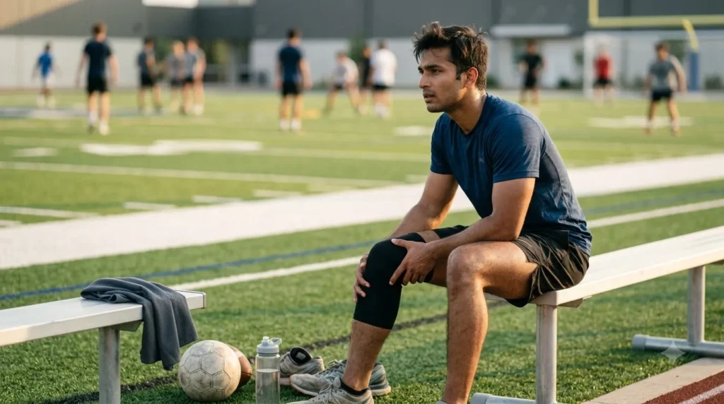
Sports Injury or Something Worse? Here’s How to Tell the Difference
There is a culture in sport, at every level, that treats pain as something to push through. The twisted ankle gets strapped and walked off. The knee that swelled after a collision gets iced overnight and tested in the morning. The shoulder that popped during a throw gets rested for a few days and then gingerly returned to. Most of the time, this approach doesn’t cause serious lasting harm. But sometimes it does – and the problem is that in the moment, it is genuinely difficult to tell which situation you are in.
The gap between a manageable sports injury and something that needs urgent medical attention is not always as obvious as people assume. A stress fracture can feel almost identical to shin splints for weeks. A partial ACL tear can allow someone to walk and even jog while the structural damage progresses. Compartment syndrome – a genuine surgical emergency – begins with muscle pain and tightness that easily resembles a straightforward muscle injury.
This post is about reading the signals more accurately – understanding which features of a sports injury suggest something manageable and which suggest something that needs professional assessment before the next training session, not after.
Every sports injury sits somewhere on a spectrum from minor tissue stress to structural damage to genuine emergency. Where an injury lands on that spectrum depends less on how much it hurts – pain is a surprisingly unreliable severity indicator in acute injury – and more on a combination of factors:
How the injury happened is one of the most diagnostically useful pieces of information. A contact tackle that hyperextended the knee with a pop heard or felt is a very different injury than a gradual knee ache that developed over several weeks of increased training. A fall on an outstretched hand with sudden wrist pain carries different implications than wrist soreness from repetitive racket strokes. High-energy mechanisms – falls from height, direct impacts, sudden forced joint movement beyond its normal range – significantly raise the probability of fracture or ligament rupture compared to gradual onset injuries.
Swelling is one of the more informative early signs. Swelling that appears within the first one to two hours of an injury and is significant – visibly distorting the joint contour – often indicates intra-articular bleeding (haemarthrosis), which in the knee is strongly associated with significant structural injury including ACL tears, fractures, or meniscal tears with vascular involvement. Swelling that develops more gradually over twelve to twenty-four hours is more typical of the inflammatory response to a sprain or muscle injury.
Bony swelling – a firm, localised prominence over a bone after impact – is more concerning than soft tissue swelling and raises fracture as a possibility. Diffuse soft tissue swelling around a joint after a twisting injury is more consistent with ligament or capsular damage.
The Ottawa Rules – validated clinical decision tools used by emergency physicians worldwide – are built around the ability to weight-bear after an ankle or knee injury. A person who cannot bear weight on the affected limb immediately after injury and at assessment is at significantly higher risk of fracture than one who can, even if both are in pain. This is not a perfect rule – some fractures do allow weight-bearing, and some severe sprains do not – but the inability to take four steps immediately after an injury is a consistent red flag for fracture.
An audible or palpable pop at the moment of injury is a clinically significant finding. In the knee, a pop with the mechanism of sudden deceleration, pivoting, or hyperextension is strongly associated with ACL rupture until proven otherwise. A pop in the ankle with inversion is associated with complete lateral ligament rupture. In the shoulder, a pop with forced abduction and external rotation raises the possibility of labral tear or shoulder dislocation. Joint sounds during movement (clicking, grinding) that were present before the injury are less significant than a single sudden pop at the moment of trauma.
Ankle sprains are among the most common sports injuries globally. The lateral ankle – the outer side – is the most frequently affected, typically from an inversion mechanism (the foot rolling inward). The anterior talofibular ligament (ATFL) is the first to sprain, and in severe sprains the calcaneofibular ligament (CFL) is also affected.
The features that raise fracture as a possibility in an ankle injury:
Without these features, the injury is more likely a sprain and can be initially managed with RICE (Rest, Ice, Compression, Elevation). With any of these features, an X-ray is warranted before returning to activity.
The knee is the most commonly injured joint in sport, and the range of possible injuries spans from minor collateral ligament sprains to complete ACL ruptures to complex meniscal tears. The mechanism tells a significant part of the story:
A knee that fills with fluid within one to two hours of injury, cannot be fully straightened or bent, feels unstable when trying to weight-bear, or has localised joint line tenderness – needs orthopaedic assessment before further activity. The inability to straight-leg raise after a knee injury (lifting the straight leg while lying down) suggests quadriceps mechanism disruption – patellar fracture or tendon rupture – which requires prompt surgical referral.
The shoulder is inherently the most mobile and therefore the least inherently stable of the major joints. Sports injuries to the shoulder range from acromioclavicular (AC) joint sprains (the bony prominence on top of the shoulder is tender and sometimes visibly prominent) to glenohumeral dislocation (the ball of the joint comes out of the socket – a visible deformity, extreme pain, inability to move the arm).
A shoulder dislocation is a medical emergency. The arm sits in an abnormal position – often held away from the body and supported by the other arm. It must be reduced (relocated) by a trained clinician, not by non-medical personnel, and should be assessed for fracture before and after reduction. Attempting to reduce a dislocated shoulder without imaging and trained personnel risks fracture, neurovascular injury, and incomplete reduction.
Rotator cuff tears from sports injury produce pain on attempting overhead or rotational movement, weakness in specific directions, and in complete tears, an inability to maintain the arm at shoulder height against gravity. Partial tears may allow function with pain. Both warrant imaging (MRI) and orthopaedic assessment, as the management decision – physiotherapy vs. surgical repair – depends on the specific tendon, the degree of tear, and the patient’s functional requirements.
Muscle strains range from mild (grade 1 – overstretching without significant fibre disruption) to moderate (grade 2 – partial fibre tear) to severe (grade 3 – complete rupture). The distinction matters because grade 3 tears of significant muscles – the Achilles tendon, the quadriceps tendon, the patellar tendon, the biceps – often require surgical repair to restore function.
Features suggesting complete rupture rather than strain:
A stress fracture is a small crack in a bone caused by repetitive loading rather than a single traumatic event. It is one of the most commonly missed diagnoses in athletes because the onset is gradual and the pain initially improves with rest, mimicking muscle soreness or tendinopathy.
The distinguishing features: pain that is consistently in the same specific location on the bone (not diffuse), that gets worse with activity and improves with rest, and that is tender to direct pressure on the bone surface rather than around it. Common sites are the tibia (shin), the metatarsals (foot bones), the navicular, and the femoral neck. The femoral neck stress fracture is the most dangerous – it can progress to a complete fracture with minimal warning, which in an older patient or a runner with bone density issues is a serious event requiring surgery. Any runner or repetitive loading athlete with groin or hip pain that is consistently activity-related and reproducible warrants imaging.
Plain X-rays often miss stress fractures in the first two to four weeks. MRI is the gold standard investigation for suspected stress fracture when X-ray is negative but clinical suspicion remains.
Compartment syndrome is a surgical emergency that develops when pressure inside a closed muscle compartment rises to a level that compromises blood flow to the muscles and nerves within it. In sports, it most commonly occurs as acute compartment syndrome following a significant direct blow, crush injury, or fracture – though it can also develop in the absence of fracture after severe muscle contusion.
The five P’s of compartment syndrome: Pain (out of proportion to the injury and worsening despite rest and elevation), Pressure (a tense, woody feeling in the affected compartment), Paraesthesia (tingling or numbness in the distribution of the nerve running through the compartment), Paralysis (weakness or loss of movement in muscles supplied by the affected compartment), and Pallor (reduced skin colour and warmth in severe cases). Not all five will be present simultaneously in early compartment syndrome, but any combination of severe increasing pain with tense compartment and sensory changes after a sports injury requires emergency assessment.
The treatment is emergency fasciotomy – surgical incision of the compartment fascia to release the pressure. The window for intervention before permanent muscle and nerve damage occurs is approximately six hours from onset of critical ischaemia. This is why compartment syndrome that is managed as a muscle bruise at home until the next morning is the injury that produces the worst outcomes.
Concussion is a traumatic brain injury caused by a direct or indirect blow to the head that produces transient neurological dysfunction. It does not require loss of consciousness – the majority of concussions involve no loss of consciousness at all – which is why it is so frequently underrecognised and undertreated in sport.
Signs of concussion: headache, dizziness, feeling ‘foggy’ or slowed, difficulty concentrating, memory gaps around the injury event, visual disturbance, light and noise sensitivity, nausea. Any athlete showing these signs after a head impact should be removed from play immediately. The ‘return to play’ principle – the idea that a concussed athlete can be assessed and returned to the same game if they seem okay – is no longer supported by evidence and is specifically contraindicated by current sports medicine guidelines.
More concerning signs that suggest intracranial haemorrhage rather than simple concussion: deteriorating level of consciousness after initial lucid period (‘talk and die’ pattern), severe headache worsening progressively, repeated vomiting, unequal pupils, or seizures. These require emergency assessment by ambulance, not a drive to the clinic.
Any sport involving contact, high speeds, or falls carries a risk of spinal injury. The critical principle: any athlete who has sustained a significant head or neck impact and is complaining of neck pain, has tingling or numbness in the arms or legs, or has any weakness after the injury should be treated as having a spinal injury until proven otherwise. This means immobilisation of the spine before moving the athlete, not examination on the field followed by a walk to the sideline. Moving an athlete with an unstable cervical spine injury without immobilisation risks converting an incomplete spinal cord injury to a complete one.
These are the sports injuries most consistently played through, most consistently undertreated, and most consistently productive of long-term problems when they are:
Go to emergency immediately
See an orthopaedic specialist within 24 to 48 hours
RICE and monitor for 24 to 48 hours
Sayee Specialty Hospital’s Orthopaedics department provides specialist consultations for sports injuries, fractures, ligament and tendon injuries, and all musculoskeletal trauma – with in-house digital X-ray and CT scan facilities for accurate diagnosis on the same visit. Whether your injury happened on the field yesterday or has been grumbling for weeks without proper assessment, our orthopaedic team provides a clear diagnosis and a management plan that actually fits your activity goals.
Playing through the wrong injury is one of the most reliable ways to turn a six-week problem into a six-month one. Getting the right diagnosis early is the fastest route back to what you want to be doing.
Book an orthopaedic consultation at Sayee Specialty Hospital, or call us at 9 976 976 976. Serving Padappai, Vandalur, Chromepet, Urapakkam, and South Chennai.
Recent Posts
Lower Back Pain Causes, Diagnosis, and Treatment Almost...

Strep Throat: Symptoms, Diagnosis, and Treatment Sore throats...

Understanding Menorrhagia: Causes, Diagnosis, and Treatment Most women...

Your Posture Is Destroying Your Spine Before 40...